Thriving Post-Surgery, Woman Finds ‘There Is Life After a Brain Aneurysm’
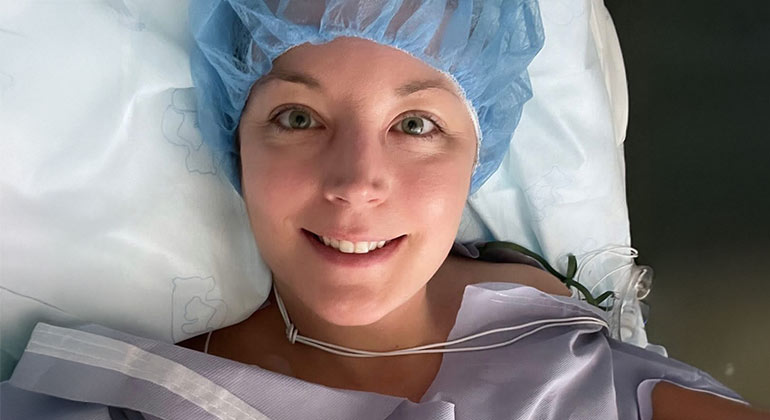
Five days after 37-year-old Julie Brothers was released from the hospital for brain aneurysm surgery, she got on a plane for Hilton Head Island. She couldn’t have imagined that scenario a few weeks earlier, when she was in an ambulance rushing to The Mount Sinai Hospital. Christopher Kellner, MD, the leading neurosurgeon who treated her, “did a great job surgically,” Julie says. “And at the same time, my body did a great job healing.”
On April 23, 2024, Julie was working from home for her job in TV production. Suddenly, she got a blinding headache. “To say it was the worst headache of my life is to downplay it,” she explains. “I felt like something had snapped inside my head.” Her initial thought was that she was having her first migraine. In addition to the headache, Julie’s vision was blurry, her neck was so stiff she couldn’t move it, and she was so nauseated that she couldn’t tolerate even a sip of water.
Eventually, Julie decided she needed some relief, so she went to her local urgent care office. They agreed it was probably a migraine and gave her a shot of high-dose nonsteroidal anti-inflammatory drug (NSAID), and a prescription for anti-nausea medication. But the pain grew even worse, and as the nausea continued, Julie started to worry about dehydration. A day and a half after her trip to urgent care, Julie went to the emergency room at Mount Sinai Morningside. “I was so blasé, I took an Uber to get there,” she said.
Scans show a ruptured aneurysm
The ER doctors listened to her symptoms and performed blood work, a urine test, and a computed tomography (CT) scan of her brain. This test uses X-rays to provide multiple views of the brain. The team diagnosed a ruptured brain aneurysm (when an enlarged artery ruptures and leaks blood into the brain) and sent Julie in an ambulance to The Mount Sinai Hospital, which has advanced facilities, equipment, and specialists to treat brain aneurysms. Julie was terrified. “I had always equated an aneurysm with an obituary,” she says.
Aneurysms are more common than many people think. Two percent of Americans have an unruptured brain aneurysm—6.8 million Americans, according to the Brain Aneurysm Foundation. About 30,000 aneurysms rupture every year, the foundation says. Half of those people die, and the remaining two-thirds end up with permanent disability, many unable to return to their previous lives. Fortunately, Julie was the outlier.
When she arrived at The Mount Sinai Hospital, the emergency room staff performed a CT scan with medical dye, called a CT angiogram. This involved threading a very narrow tube called a microcatheter up through Julie’s thigh to her brain and injecting medical dye. This test showed the exact location of the aneurysm. “The blood pattern was very characteristic of aneurysm bleeding,” says Dr. Kellner, Director of the Intracerebral Hemorrhage Program at Mount Sinai.
Advanced procedure with coils
Dr. Kellner treated the aneurysm with coil embolization. Through the catheter in Julie’s leg, he delivered several tiny platinum coils into the aneurysm. The coils entered the aneurysm straight as a pin and then Dr. Kellner carefully manipulated them into different coiled shapes, filling the space inside the aneurysm so it could not collect blood again.
“Fortunately, Julie’s aneurysm was shaped like a lollipop, with a thin neck leading to a round area. The narrow neck of the aneurysm helps keep the coils in and the blood out,” Dr. Kellner explains. The procedure took less than two hours, and then Julie spent 15 days in the neurology intensive care unit (ICU).
“Blood in the brain can be very serious; patients have a one in five chance of dying. And it can also cause a variety of other problems,” Dr. Kellner says. “Some people need an additional procedure after the first one.” Fortunately, Julie did not have any of those issues.
In the ICU, the medical staff started right away to get Julie back up to speed. “On the second day, they started getting me to eat lunch sitting up, then they progressed to having me sit in a chair for every meal. Then they had me sit in a chair for an hour every day, then three hours a day,” Julie says. She started walking to the bathroom, though the ICU rules were very strict, and she was always accompanied by a nurse because she was at risk of falling.
“The nurses woke me up every hour through the night to make sure I hadn’t lost any physical or neurological abilities. Fortunately, all my fingers and toes were working, and there was no sign of neurological problems,” Julie says. “I was one of the lucky ones,” Julie moved to a step-down room for three days. “They took out multiple IVs, and I could finally take a shower.”
Julie’s parents drove in from Ohio as soon as they heard the news. They stayed in her apartment and looked after her cat while she was in the hospital, and visited her twice a day. “Dr. Kellner took plenty of time to talk with me and my parents and answer all our questions,” Julie says.
Julie appreciated the treatment she got from everyone at The Mount Sinai Hospital. “I got excellent care, from the surgeon to the nursing staff to everyone else in the hospital.” She even enjoyed the hospital food. “I posted my meals on Facebook and Instagram.”
“He was very patient and helpful”
By the time Julie was discharged, she was able to do everything she could do prior to the aneurysm. Before she left, she talked with Dr. Kellner. “I had a list of 32 questions, and he answered every one; he was very patient and helpful. He really cared about me.” Julie received medication to manage her pain, to help her sleep, and to keep her blood pressure down. Her parents stayed in New York to help for a few days after she got home.
Five days after Julie was discharged, she flew to Hilton Head Island for a long-planned family vacation. “I asked Dr. Kellner if it was safe for me to fly, then to go swimming and biking. He said yes, I didn’t have to hide in my room,” Julie says. “I love to travel, and it was great to know that I could.”
Six months after the procedure, Dr. Kellner did a follow-up angiogram. The imaging test showed that the aneurysm remained completely treated, with no blood flow to the aneurysm and no additional aneurysms. “I felt like I got an ‘A’ on the test,” Julie says.
Confident in her ability to do her job well, Julie went back to work. Even so, she will need periodic magnetic resonance imaging (MRI) tests of her brain, because once someone has had an aneurysm, their arteries are prone to forming future aneurysms. “We want to check periodically, just in case. We use MRIs to cut down on the radiation that she would get with CT scans,” Dr. Kellner says. Since the follow-up was clean, Julie’s next imaging test will be in 2027. “If that looks good, we’ll check her every five years,” Dr. Kellner says.
Julie is back to her pre-aneurysm life. “I have a little fatigue and a little brain fog. I’d say I’m 99 percent back to where I was.” Four months after the procedure, she participated in the 5K walk/run. “I saw Dr. Kellner and got to meet his wife and kids, who were there to cheer him on. It was the best!” Julie continues to raise money for the Brain Aneurysm Foundation, which funds research into the disease. “I feel very lucky and want to do everything I can to get the word out,” Julie says. “There is life after a brain aneurysm.”