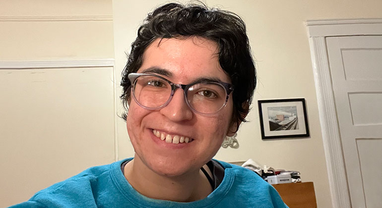
Innovative Surgery Treats A Young Woman With a Ruptured AVM, a Tangle of Vessels in the Brain
In December 2021, Claudia Mallea, a photo-archivist and digital curator, was looking forward to celebrating her 24th birthday with her roommate in their Brooklyn apartment. She was recovering from COVID-19 and had boiled some water to fill her neti pot to help clear her sinuses. When she got to the bathroom, she collapsed. “I carried the kettle into the bathroom, set it down in the sink, and started to feel odd,” Claudia says. “Then I lost my balance, fell to the floor, and became paralyzed down my left side.”
She managed to drag herself back into the kitchen, where her roommate found her, but she was unable to stand and started to have seizures. Claudia remembers very little from that point on, but learned later that her roommate called an ambulance and that the paramedics recognized from her slurred speech and paralysis that she was having a stroke.
This episode was the beginning of a long and complex medical journey that would lead Claudia to receive highly specialized treatment and care at The Mount Sinai Hospital for a life-threatening condition related to a blood vessel malformation in her brain.
The paramedics immediately took her to the nearest hospital, where she underwent brain and body imaging that led to her diagnosis—a ruptured arteriovenous malformation (AVM) in her brain.
Arteries carry blood from the heart to the brain, where blood vessels called capillaries extract the oxygen and nutrients. The blood then returns to the heart through veins. With AVM, an abnormal tangle of blood vessels prematurely connects the arteries directly to the veins, bypassing the normal capillary circulation. Such malformations happen during the development of the brain before birth, and they can rupture, causing bleeding within the brain that can lead to injury or even death.
The hospital medically stabilized her, and on December 29—her 24th birthday—she was transferred to The Mount Sinai Hospital for advanced treatment. On the same day, Claudia first underwent a hemicraniectomy (where a section of the skull is removed) to reduce the pressure on her brain caused by the bleeding. This was performed by neurosurgeon Christopher Kellner, MD, Director of Mount Sinai’s Intracerebral Hemorrhage Program. This was immediately followed by an endovascular embolization procedure carried out by Hazem Shoirah, MD, Director of Thrombectomy Services at Mount Sinai Queens, to stabilize the ruptured AVM. During this procedure, he found that the AVM had two aneurysms, which were the cause of her hemorrhage. These aneurysms were treated by embolization—a technique where aneurysms are closed up with a glue-like material through a small catheter. The embolization significantly decreased the risk of another immediate rupture and allowed for future elective embolization procedures to take place.
Claudia spent a month in rehabilitation, before returning to Dr. Kellner in February 2022 for a craniosplasty. The procedure involved attaching a prosthetic piece of skull to the area where bone had been removed during the earlier hemicraniectomy. She then spent another month in rehabilitation.
Treatment of the ruptured AVM was then overseen by Dr. Shoirah. Over the next three months, Claudia had periods of rest at home and rehab as Dr. Shoirah performed a series of embolization procedures to further stabilize and shrink the ruptured AVM.
At the end of May, she returned one more time to The Mount Sinai Hospital for her final procedure—a new technique called transvenous embolization. Dr. Shoirah was assisted by Alejandro Berenstein, MD, Director of the Pediatric Cerebrovascular Program, Johana T. Fifi, MD, Associate Director of the Cerebrovascular Center, and visiting professor René Chapot, MD, a renowned expert in the procedure.
“Transvenous embolization is a new, advanced technique,” Dr. Shoirah explains. “Specially designed microcatheters are used to precisely navigate through the veins, to reach the one vein that is draining the AVM. The draining vein is plugged with coils, and a liquid material is then injected from the draining vein into the tangle of blood, closing up and curing the AVM. This technique offers the advantages of being minimally invasive and highly effective, and can achieve a complete cure in a single session.”
The surgery was a success, and there has been no further blood flow into the AVM. “Thanks to the cutting-edge treatment available to me as a Mount Sinai patient, I was able to have my AVM cured without further compromising my neurological function,” Claudia says.
The path to recovery remains a long one, and she continues to receive long-term rehabilitation care and to work on improving her mobility and cognitive skills.
“I am very much in the process of recovering, but very relieved to have my AVM treated,” Claudia says. “My mobility has improved a lot through therapy, but I still have a slow walking speed and use a cane and an ankle foot orthoses to stabilize my ankle. I have limited use of my left arm, and continue to experience some minor cognitive issues. I do physical therapy twice a week and cognitive therapy weekly. I feel proud of the progress I've made so far and am relieved to no longer have something at risk of rupture in my brain.”