
Obtaining Prefrontal Cortex Biopsies During Deep Brain Stimulation Adds No Risk to Procedure
Mount Sinai findings address unmet need for collecting living human brain tissue to advance medical research
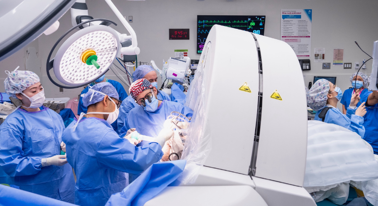
Obtaining prefrontal cortex biopsies during deep brain stimulation (DBS) surgery in living patients does not increase the risk of adverse events or cognitive decline compared to standard DBS procedures that don’t involve biopsies, a team of clinical research scientists at the Icahn School of Medicine at Mount Sinai has demonstrated.
The study findings, published online September 3 in Neurosurgery, the official publication of the Congress of Neurological Surgeons, establish the safety of an approach that allows researchers to collect valuable living human brain tissue during planned neurosurgical procedures, addressing a critical barrier in neuroscience research while maintaining patient safety. Most studies of how the brain works at the molecular level have been performed using samples from deceased patients because of the lack of safe approaches to sampling the living human brain.
“Through the Living Brain Project at Mount Sinai, we developed a method to safely biopsy a small volume of brain tissue from a region of the prefrontal cortex during placement of the deep brain stimulation lead,” said Brian Kopell, MD, Director of the Center for Neuromodulation, co-lead of the Living Brain Project at Mount Sinai, and co-senior author of the paper. “The retrospective safety analysis clearly demonstrates that there are safe and innovative ways of involving neurosurgical patients in research activities that have the potential to greatly advance science. We extend our deepest gratitude to the patients who have chosen to partner with us on this work.”
The research team analyzed acute adverse events, defined as infection, intracranial hemorrhage (ICH), and seizures, following 1,152 deep brain stimulation lead placements performed on 590 patients at a single hospital between 2013 and 2024. Prefrontal cortex biopsies were obtained in 652 procedures (“biopsy group”) and not obtained in 500 procedures (“non-biopsy group”). A computed tomography (CT) scan was performed within hours of each procedure and patient medical records were reviewed for acute ICH, seizure, and infection from the procedure date to 90 days later. A subset of patients was followed for approximately one year to assess cognitive outcomes.
No infections occurred in either group. No statistically significant differences in ICH rate or seizure rate were observed between groups (ICH rate: 1.7% biopsy group vs. 1.4% non-biopsy group; chi-square test p-value=0.88; seizure rate: 0.2% biopsy group vs. 0.4% non-biopsy group; p-value=0.82). No statistically significant associations were observed between number of biopsies and changes in cognitive health over time.

“The ability to safely study the brain from people who are alive opens up a new world of questions about brain function that researchers can tackle,” said Alexander W. Charney, MD, PhD, Director of The Charles Bronfman Institute for Personalized Medicine, co-lead of the Living Brain Project, and co-senior author of the study. “By using this technique, the Living Brain Project is already making several contributions to medical research, including a comprehensive characterization of living human brain biology, technology to study human brain circuits at the synapse level, technology to deliver gene therapy to the brain, and insights into the pathogenesis of brain disease. Given the safety profile of the technique, we are eager for other institutions to follow the same methods and expand the scope of this work.”
Deep brain stimulation is an elective neurosurgical treatment for neurological and mental health illnesses. A common technique for safely implanting the DBS electrode involves cauterizing a small volume of prefrontal cortex prior to inserting the cannula, a specialized hollow tube through which the electrode lead is placed. For the current study, all DBS procedures were performed by a single neurosurgeon using standard stereotactic techniques. All procedures involved a standard frontal burr hole and stereotactic cannula placement, with cortical surface preparation differing only by the inclusion of a prefrontal cortex biopsy (obtained prior to cauterization and using a standard punch tool). Biopsy sizes were measured for 231 biopsies. The mean biopsy volume measured was equal to 40mm3 and the median volume was equal to 30mm3. The study authors note that the tight biopsy sizes distributed around the mean demonstrate that the Living Brain Project prefrontal cortex biopsy procedure can be highly standardized, reproducible, and precise.
“The innovative approach that the Living Brain Project team carefully and meticulously developed has allowed analysis of living human brain tissue for the first time, transforming our capability to study the function of the human brain and providing new insights into the causes and treatment of brain diseases,” said Eric J. Nestler, MD, PhD, Interim Dean and Nash Family Professor of Neuroscience at the Icahn School of Medicine at Mount Sinai; Executive Vice President and Chief Scientific Officer of the Mount Sinai Health System. “With more than a decade of data that demonstrate exemplary safety standards, the Living Brain Project is paving the way for other researchers to follow suit and help advance science beyond what has ever been possible before.”
About The Living Brain Project
The Living Brain Project is a multiscale, data-driven investigation of the living human brain that uses the full human subjects neuroscience toolkit to discover how interactions between the various levels of neurobiology, neurophysiology, and neuroanatomy give rise to neuropsychiatric functioning. The toolkit includes clinical observation, neuropharmacology, neuroimaging, neurophysiology, neuromodulation, and molecular-cellular neuroscience. Led by Drs. Charney and Kopell, the study has enrolled more than 600 individuals undergoing elective deep brain stimulation surgery for neurological and neuropsychiatric conditions including Parkinson’s disease, obsessive-compulsive disorder, essential tremor, dystonia, and depression. The project has received funding from the National Institute of Health’s National Institute on Aging.
About the Mount Sinai Health System
Mount Sinai Health System is one of the nation’s leading integrated academic health systems and one of the largest in the New York metropolitan area. Its comprehensive system includes seven hospitals, more than 400 outpatient practices, over 600 research and clinical laboratories, the Icahn School of Medicine at Mount Sinai, the Graduate School of Biomedical Sciences, and the Mount Sinai Phillips School of Nursing. Together, the Health System comprises approximately 48,000 employees, more than 9,000 physicians, and 8,600 nurses.
As a leading learning health system, Mount Sinai combines clinical expertise with scientific discovery to improve patient care while training the next generation of health care and biomedical leaders. The Health System provides care across every stage of life, from prenatal care through geriatrics, while advancing personalized medicine through artificial intelligence, data science, and biomedical research.
Mount Sinai is consistently recognized among the nation’s leading academic health systems for patient care, research, and education. The Mount Sinai Hospital is ranked No. 1 in New York by Newsweek and No. 5 on the magazine’s World’s Best Hospitals list. The Icahn School of Medicine at Mount Sinai ranks No. 11 among U.S. medical schools and No. 1 among freestanding medical schools for National Institutes of Health funding, reflecting the strength of its scientific enterprise and leadership in biomedical research.