Brain Scan-Blood Test Panel Promises Improved Diagnosis of Brain Trauma Following Battlefield Blast Exposure
New brain scans and blood tests move researchers towards more sensitive diagnosis of battlefield brain trauma and evaluation of new drugs
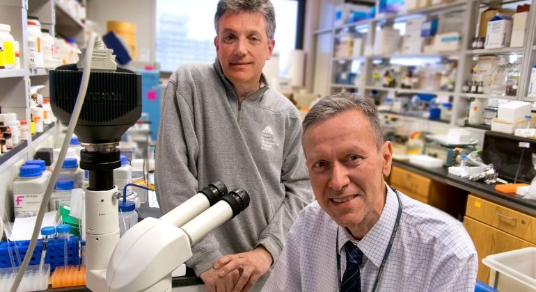
Samuel Gandy, MD, PhD, and Gregory Elder, MD in the lab at James J. Peter VA Medical Center.
An array of tests that combines functional assessment with blood tests and brain scans promises more sensitive and objective estimation of brain degeneration in human veterans exposed to battlefield improvised explosive device (IED) blasts, according to research led by doctors at the Icahn School of Medicine at Mount Sinai and the James J. Peters VA Medical Center. The study was published in Molecular Psychiatry on Tuesday, February 25, and featured on the journal’s cover.
Traumatic brain injury (TBI) is associated with acute brain destruction at the time of the injury and is also a risk factor for developing neurodegenerative diseases later in life. It is estimated that 10 to 20 percent of veterans returning from the conflicts in Iraq and Afghanistan sustained mild TBI resulting from IED and other blast exposures. The true prevalence may be even higher, given that many blast-related injuries go undocumented. Symptoms of mild TBI frequently resolve in days to months following injury, but in a subset of patients, symptoms persist and evolve into a chronic syndrome. Veterans who had sustained TBIs may suffer subtle, yet important, endocrine and neuropsychiatric dysfunction. Given the fact that suicide rates have jumped substantially among young military veterans in recent years, it is imperative to develop earlier, more objective and sensitive methods to detect brain damage.
Using advanced methods of clinical neuropsychological and neurocognitive assessment, brain imaging, and blood biomarker measurement in veterans of the Middle East conflicts, together with modeling of mild repeated blast injury in laboratory rats, the researchers found changes in the brains and blood of those subjected to blasts. Specifically, they performed neuroimaging and blood analysis of human veterans who were exposed to IED blasts on the battlefield and on rats exposed to repetitive, low-level blasts in a shock tube. All veterans reported histories of between 1 and 50 blast exposures, and all had chronic behavioral and cognitive complaints.
Two chemical changes that occur during neurodegeneration involve clumping of a brain protein called tau and leakage of another protein called neurofilament protein-light chain (Nf-L) from the brain into the blood. The research team used positron emission topography (PET) and the [18F]AV1451 (flortaucipir) tau ligand, a molecule which produces a signal by binding to a site on the tau protein that “lights up” on a PET image, and found that 5 of the 10 veterans exhibited excessive retention of [18F]AV1451 at the white/gray matter junction in frontal, parietal, and temporal brain regions, a typical localization where tau protein clumps tend to accumulate after TBI. In healthy brains, tau is essential for normal cell functioning, helping stabilize the internal skeleton of nerve cells in the brain, but when tau proteins build up and clump together, it causes the internal skeleton to collapse and form twisted tau tangles that promote brain cell damage.
As an additional biomarker, they measured blood levels of Nf-L, since elevated levels of Nf-L have been reported in human patients suffering from a variety of brain injuries, including mild TBI and neurodegenerative diseases. They observed elevated levels of Nf-L in the plasma of veterans displaying excess [18F]AV1451 retention. The human component of the study was led by Sam Gandy, MD, PhD, Professor of Neurology, and Psychiatry, and Director of the Center for Cognitive Health and NFL Neurological Care at the Icahn School of Medicine at Mount Sinai, Attending Neurologist at the James J. Peters VA Medical Center, and co-senior author of the paper.
In parallel, the research team examined a rat model being studied by Gregory Elder, MD, Professor of Neurology, and Psychiatry, at the Icahn School of Medicine at Mount Sinai, Chief of Neurology at the James J. Peters VA Medical Center, and co-senior author of the paper. The rat model was designed to mimic a level of blast exposure that would be comparable to a mild TBI or a subclinical blast exposure in humans. Rats exposed to this blast protocol exhibit a range of anxiety and behavioral traits resembling post-traumatic stress disorder. Dr. Elder and colleagues found that rats exposed to repetitive, low-level blasts accumulated abnormal tau in nerve cells as well as around blood vessels in cells called astrocytes. Astrocytes play important roles in supporting nerve cells and during inflammatory events.
“We are fortunate to have access to both living humans and living rodent models so that we can conduct side-by-side comparisons of the clinical and microscopic changes that are common to both species related to traumatic brain injury,” said Dr. Gandy. “As a result of these parallel studies in veterans and in the Elder brain injury model, we are well on our way to the first clinical trials wherein first-in-class drugs will be evaluated for their safety and for their potential clinical benefit in relieving the anxiety, depression, memory disorders, and anger management issues that are associated with traumatic brain injuries.”
Previous research conducted by Dr. Gandy and his colleagues identified some of the first PET images of protein aggregates in the brains of living athletes and veterans with histories of TBI, consisting primarily of either the tau protein or, alternatively, a protein called amyloid beta. Amyloid beta is the main component of brain plaques associated with Alzheimer’s disease, while tau is the main constituent of neurofibrillary tangles, which are hallmarks of the neurodegenerative diseases frontotemporal dementia and chronic traumatic encephalopathy (CTE). For now, CTE can only definitively be diagnosed postmortem, but one goal of this research conducted by Drs. Gandy and Elder is to find methods for effective diagnosis while patients are still alive.
“There are many young, otherwise healthy veterans who have suffered blast-related TBIs, some of them years in the past, who either aren’t getting better or, in some cases, are getting worse,” says Dr. Elder. “We don’t know why or how to identify those at greatest risk. The work in this study is a step towards answering those questions.”
For this project, the human studies were conducted by Dara Dickstein, PhD, Adjunct Assistant Professor of Neuroscience at the Icahn School of Medicine at Mount Sinai.
Following blast exposure, the rats were studied by Rita De Gasperi, PhD, Instructor in Psychiatry at the Icahn School of Medicine at Mount Sinai and the James J. Peters VA Medical Center.
“This research supports the Alzheimer’s Drug Discovery Foundation mission by identifying biomarkers — like brain neuroimaging — that can lead to earlier diagnosis of Alzheimer’s and related dementias, including CTE,” said Howard Fillit, MD, Founding Executive Director and Chief Science Officer of ADDF, one of the study’s financial supporters. “CTE research is relatively new, but it is advancing rapidly. Studies such as this one will advance our understanding of CTE, Alzheimer’s and other neurogenerative diseases, improve the rigor and efficiency of clinical trials, and may ultimately provide screening tools to identify patients for the trials and as a measurement to assess drug effect.”
The study was primarily supported by the Alzheimer’s Drug Discovery Foundation and the Department of Veterans Affairs.
About the Mount Sinai Health System
Mount Sinai Health System is one of the largest academic medical systems in the New York metro area, with 48,000 employees working across seven hospitals, more than 400 outpatient practices, more than 600 research and clinical labs, a school of nursing, and a leading school of medicine and graduate education. Mount Sinai advances health for all people, everywhere, by taking on the most complex health care challenges of our time—discovering and applying new scientific learning and knowledge; developing safer, more effective treatments; educating the next generation of medical leaders and innovators; and supporting local communities by delivering high-quality care to all who need it.
Through the integration of its hospitals, labs, and schools, Mount Sinai offers comprehensive health care solutions from birth through geriatrics, leveraging innovative approaches such as artificial intelligence and informatics while keeping patients’ medical and emotional needs at the center of all treatment. The Health System includes approximately 9,000 primary and specialty care physicians and 10 free-standing joint-venture centers throughout the five boroughs of New York City, Westchester, Long Island, and Florida. Hospitals within the System are consistently ranked by Newsweek’s® “The World’s Best Smart Hospitals, Best in State Hospitals, World Best Hospitals and Best Specialty Hospitals” and by U.S. News & World Report's® “Best Hospitals” and “Best Children’s Hospitals.” The Mount Sinai Hospital is on the U.S. News & World Report® “Best Hospitals” Honor Roll for 2025-2026.
For more information, visit https://www.mountsinai.org or find Mount Sinai on Facebook, Instagram, LinkedIn, X, and YouTube.