Robotic Kidney and Reconstructive Surgery
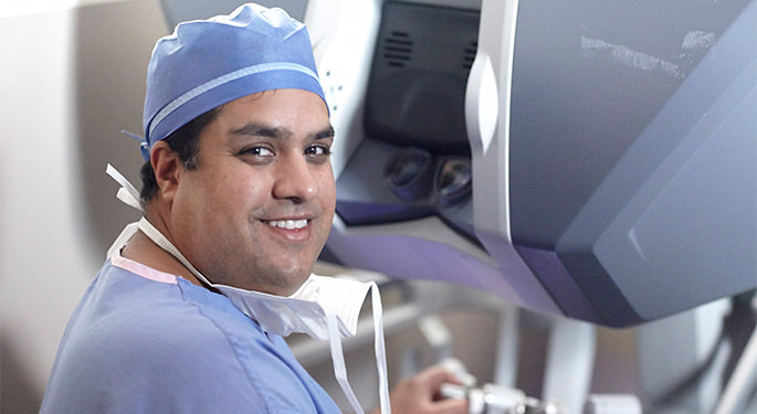
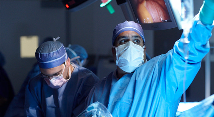
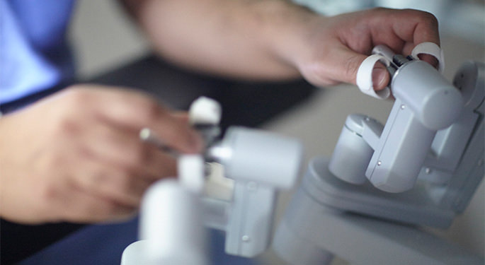
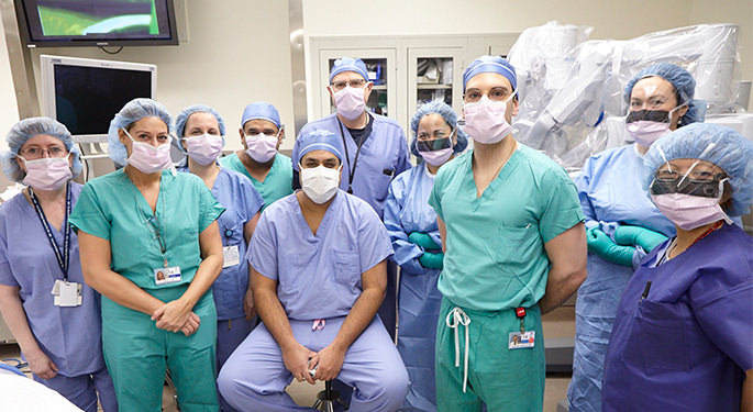
At Mount Sinai Urology, our experience with robotic kidney cancer and reconstructive surgery is extensive. Our robotic partial nephrectomy approach allows for a precise excision of the tumor while leaving the healthy and functional part of the kidney intact. In addition, minimally invasive robotic surgery offers fewer complications and side effects, smaller incisions, less scarring, decreased post-surgical pain, shorter hospital stays, and faster recovery.
F.A.S.T. Robotic Partial Nephrectomy
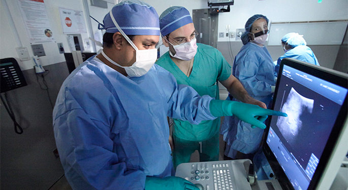
F.A.S.T. robotic partial nephrectomy is an advanced approach to surgically remove a kidney tumor and reconstruct the kidney to prevent functional damage. Developed by Ketan K. Badani, MD, Director of the Comprehensive Kidney Cancer Program, this procedure enables the surgeon to minimize the damage to the normal part of the kidney by precisely removing the tumor along using real time ultrasound mapping, and minimizing time the kidney is without bloodflow during this critical portion of a partial nephrectomy. This is known as ischemia time, and by reducing this number, we also lower the changes of kidney loss over a patient's lifetime. This approach also allows us to more safely and reliably perform partial nephrectomy (compared to removing the entire kidney) in even the most complex tumor sizes and locations while still offering the best outcome possible for the patient.
Dr. Badani originally developed F.A.S.T. as a means to shorten partial nephrectomy ischemia time. This is a critical point when the kidney is without blood flow while the tumor is being excised and the kidney reconstructed. Historically robotic surgeries involved the surgeon plus any number of assistants who are called on to carry out important portions of the operation. In a F.A.S.T. robotic partial nephrectomy, we never leave the controls. Dr. Badani's technique takes certain steps out of the hands of the assistant, and puts them back into the hands of the robotic surgeon. That shaves minutes off the procedure and reduces the amount of time in which blood flow to the kidney is stopped (ischemia time). If you have kidney cancer, that's better for your kidney, and better for you.
The F.A.S.T. technique also allows Dr. Badani to perform robotic partial nephrectomies on increasing numbers of complex kidney tumors rather than having to remove the entire kidney. It is typical for surgeons to remove the entire kidney if the tumor is large, or close to key structures in the kidney. The F.A.S.T. approach allows us to safely and effectively save kidneys, even in complex circumstances.
Benefits
The benefits of F.A.S.T. include:
- Using small “keyhole” incisions instead of a major incision results in less blood loss and faster recovery
- Decreased ischemia (by as much as 50 percent) and operative time lowers the chances of poor outcomes
- Enhanced visualization and tumor identification allows us to see both the operative field and ultrasound images simultaneously
- Immunofluorescence imaging enables us to focus on the specific branch that feeds the tumor, clamp it, and maintain blood flow to the rest of the kidney. This decreases, or even eliminates, ischemia time in many cases
Robotic Surgery for Ureteropelvic Junction (UPJ) Obstruction and Ureteral Reconstruction
Blockage of the first part of the tube (ureter) that collects urine from the kidney and drains it into the bladder can cause a backup of urine, leading to pain, infections, stones, high blood pressure—and eventually may lead to kidney damage. This ureteropelvic junction (UPJ) obstruction usually results from congenital (inherited) narrowing of the ureter where it meets the kidney. Blockage of the UPJ may also be caused by compression of a nearby blood vessel, kidney stones, inflammation, or previous surgery.
Other areas of the ureter (tube that connects the kidney to the bladder) can also become damaged for various reasons such as prior surgery, radiation, tumor, stones, and inflammation. These conditions can also be treated via a minimally invasive robotic approach using advanced technology such as intraoperative real time ultrasound, immunofluorescence vision, tissue perfusion characteristics, and state of the art biologic materials to assist in reconstructing the ureter.
Robotic-assisted Pyeloplasty
Robotic pyeloplasty is generally the most common surgery for UPJ obstruction. In pyeloplasty, we typically remove the blocked portion of the ureter and reconnect the healthy ends to re-create a free-flowing connection. A plastic stent (tube) is temporarily left inside the repaired ureter for support. It is removed in the office a few weeks later.
The robotic procedure is typically performed through 3 to 4 small incisions on the abdomen. Compared to the open pyeloplasty, robotic techniques may allow precise correction of the UPJ obstruction with smaller incisions, which may reduce blood loss, decrease pain, shorten hospital stay (usually 1 day versus several days to a week), and lead to a faster overall recovery.
The robotic technique is especially helpful in this delicate UPJ surgery as it requires a high level of precision to avoid blood vessels near the ureter and to prevent scar formation (fibrotic) tissue. This is accomplished by examining the perfusion and health of the ureter using near infrared imaging during the surgery. The enhanced optics and greater dexterity provided by the robotic system allow the experienced surgeon to accomplish all these technically demanding tasks—all performed deep inside the abdomen—precisely.
Robotic Ureteral Reconstruction
There are several types of ureteral conditions, and the approach to repair will depend on the length of damage, and the location (upper, mid, or lower ureter).
For blockages (called strictures), surgery often involves removing the blocked portion of the ureter and reconnecting the healthy ends to re-create a free-flowing tube. A stent (a plastic tube) can be inserted inside the repaired ureter for support.
If the ureteral blockage is near the bladder, we can directly reconnect the tube to the bladder called a re-implant. This can be done for ureteral damage in the bottom half of the ureter.
In a special procedure for patients with retroperitoneal fibrosis, the surgeon performs a ureterolysis—a complex procedure that frees the ureter from encasing fibrotic adhesions (scar tissue) that block the flow of urine.
The robotic approach to this condition allows for precise sewing and ensuring healthy edges of tissue are being used to prevent future scarring and obstruction. Additionally, if you are undergoing robotic surgery, you can expect a shorter hospital stay, faster return to normal activities, less pain, and lower rate of complications.
Robotic Adrenal Tumor Surgery and Simple Nephrectomy
Robotic adrenalectomy is commonly offered for most adrenal tumors or hyperfunctioning glands. Unlike traditional open adrenalectomy, which requires a large flank incision, robotic-assisted adrenalectomy is conducted through four or five tiny incisions in the abdomen. The robotic approach allows superior visualization and dexterity along with advanced instruments. These are key advantages that provide unparalleled precision to the delicate removal of adrenal tissue from the many arteries and veins that feed this small hormone-producing gland. Precision and accuracy are of utmost importance in this crowded anatomical region in order to avoid any contact with the nearby spleen, liver, pancreas, small bowels, aorta and/or the vena cava.
We also perform partial adrenalectomy, where if the tumor is not directly in the middle of the gland, we can remove only the tumor and preserve the normal part of the adrenal gland.
As with most robotic-assisted procedures, there is typically far less pain than with open procedure, less blood loss (and therefore less need for a blood transfusion), less scarring (both internally and externally), a shorter hospital stay (1 to 2 days versus up to a week), and a shorter overall recovery time before returning to normal activities.
Robotic Simple Nephrectomy
For a non-functional kidney, robotic simple nephrectomy is recommended when a diseased kidney stops working due to longstanding obstruction, kidney stones, or infection. Many times, we need to remove these kidneys before challenges arise. We perform this operation through three or four small holes and most patients are home by the next day.
Diagnosis and Treatment Options
Diagnosis
We work very closely with key members in radiology who specialize in imaging kidneys and the abdomen. This program uses high-resolution CAT scan and MRI technology to provide highly detailed images that help us determine which branches of the kidney are feeding the tumor and should be removed.
Treatment Options
Minimally invasive robotic surgery is not an option for everyone. The decision to proceed with robotic surgery depends on numerous factors, including the size, location, and aggressiveness of the tumor as well as your medical profile and history, your expectations, and preferences.
We use several other approaches to treat kidney cancer if you are not a good candidate for robotic surgery. These include ablation (cryotherapy to freeze the tumor, and radiofrequency ablation to heat the tumor), angiography and embolization, and traditional open surgery.
As the Director of the Comprehensive Kidney Cancer Program, Dr. Badani has assembled a team of leading experts as part of your multi-disciplinary treatment program including nephrology, radiology, pathology, medical oncology, and radiation oncology.
Recovery and Hospital Stay
If you undergo robotic kidney surgery, you will likely be able to go home the following day. We offer optional private rooms with concierge service at our Mount Sinai Hospital and Mount Sinai West locations. We see patients during the hospital stay and one to two weeks after surgery for a check-up and to discuss pathology results. At that point, a surveillance program to monitor post treatment will be formulated. If you are interested in a private room, please call the office at 212-241-3919.
It usually takes half as long to recover from robotic kidney surgery as from an open procedure. You can typically return to work one to four weeks later, depending on how physically demanding your job is. Most people go back to work and normal activities within three weeks of the procedure.
We serve you a light dinner the night of surgery and you should be eating normally within a day or two after the procedure. We encourage you to start doing moderate and non-resistance exercises such as walking, jogging, and swimming two weeks after the procedure. We do NOT recommend lifting weights until three to four weeks following surgery.
Surveillance
Three to six months after the surgery, we conduct imaging tests to assess your kidneys and the surrounding areas. We may use a computed tomography (CT Scan) or magnetic resonance imaging (MRI) scan and/or a chest x-ray. How often we see you will depend on tumor pathology (i.e., whether it was benign or malignant and how aggressive it was growing). Generally, you will come in twice a year for blood tests and once annually for imaging. If you traveled to Mount Sinai for the surgery from out of state or the country, you can have follow up tests performed near your home. We will keep in touch with your personal physician to ensure follow up is seamless and up-to-date.
Holistic Kidney Treatment
A key member of the Comprehensive Kidney Cancer Program is Jillian Capodice, MS, LAC, Assistant Professor and Director of the Integrative Urology and Wellness Program at Mount Sinai. Jillian develops customized nutrition, exercise, and stress management programs to facilitate post-surgery recovery and help you cope with side effects and maintain the highest degree of wellness moving forward. She can help formulate a diet and nutrition plan that is “kidney healthy” after surgery. As obesity and high blood pressure are risk factors for kidney cancer, she can also consult with you about lifestyle changes and disease management strategies.

