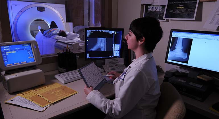
Musculoskeletal Imaging
We combine the latest technology with the most advanced clinical expertise to diagnose and treat conditions in your bones, joints, and soft tissues. We work closely with experts in orthopedics, rheumatology, rehabilitation medicine, and other specialties. We also partner with your referring physician to deliver the most accurate diagnosis and deliver the best possible outcome.
We use a variety of imaging tests, such as ultrasound and magnetic resonance imaging (MRI). We also use special imaging tests that are specifically designed for musculoskeletal conditions. These include:
- Musculoskeletal Ultrasound Imaging uses high-frequency sound waves to evaluate bones, joints, tendons, and ligaments. It does not use ionizing radiation (X-rays).
- X-Ray (Radiography) is the most basic imaging test used to evaluate possible abnormalities or trauma to the musculoskeletal system. It provides a cursory look at the bones, joints, and soft tissues and is the first step in the assessment of abnormalities in the musculoskeletal system.
- MRI (Magnetic Resonance Imaging) uses a large magnet to produce detailed evaluation of the structures of the musculoskeletal system. MRI provides great anatomic detail of the structures around joints, such as tendons, ligaments, labrum, menisci, and cartilage. This modality also offers detailed evaluations of bone marrow in cases of tumor or infection, as well as the supporting muscles and nerves.
In addition to standard MRI imaging, we use advanced technologies such as the GE Extremity MRI, which is an open system dedicated to scanning hands, wrists, elbows, feet, ankles, and knees with the greatest level of comfort and convenience.
- MR Arthrography provides fine image resolution of various joints, usually the hip or shoulder. Your doctor may order an MR arthrogram for a more detailed evaluation than a standard MRI. This test requires the injection of diluted MRI contrast into the joint. Then we perform the MRI. The presence of contrast allows for detailed evaluation of the joint, particularly the cartilage and labrum. MR arthrography also helps detect subtle tendon tears and is useful if you have already had surgery.
- CT (Computed Tomography) scans provide a more detailed evaluation of the bones, joints, and soft tissues than x-ray. We use CT scans to obtain detailed evaluation of fractures, bone architecture, and orthopedic hardware. CT scans can also give us three-dimensional images.
Treatments We Offer
Not only do we use imaging for diagnoses, but we also perform a number of therapeutic procedures. The procedures we perform most often are:
- Bone and Soft Tissue Biopsy. We use a combination of light sedation and local anesthesia to perform bone biopsies, guided by a CT scan. We use either ultrasound or CT scans to guide soft-tissue biopsies.
- Calcific Tendinitis Injection. Calcific tendinitis is caused by the deposition of calcium in and around tendons and joints. It can occur anywhere in the body but most often around the shoulders and hips. This condition is extremely uncomfortable, and the inflammation makes it hard to move. We find that ultrasound-guided injection is very effective. Once we pinpoint where the calcific deposits are, we numb the larger area with local anesthesia. Then we inject the calcium deposit with anesthesia and use a needle guided by ultrasound to dissolve the calcium in and around the tendon. Afterwards, we inject a steroid into the tendon and bursa to reduce inflammation and promote healing.
- Morton's Neuroma Injection. Morton's neuroma is a benign lesion that occurs in the feet, usually between the toes. It appears most often between the third and fourth toe. The condition can cause a sharp, burning pain. It is usually the result of scarring around the small nerves between your toes. Usually, we first treat these conditions conservatively, by recommending you change footwear and use arch supports. If this doesn’t work, we inject local anesthesia and a steroid. We use ultrasound imaging to pinpoint the precise spot for the injection, which usually reduces your pain and inflammation. If it does not work, we can use the same procedure to inject the area with alcohol to kill the surrounding inflammatory tissue.
- Radiofrequency Ablation of Osteoid Osteoma. Osteoid osteoma is a benign bone tumor found mostly in young adults. We usually find this condition when a patient reports pain in the arms or legs. Usually this pain is more frequent or severe at night and goes away with aspirin or ibuprofen. This can be a complicated procedure and sometimes requires a long hospital stay. Instead, we often use radiofrequency ablation, which involves applying intense heat to the tumor, guided by a CT scan. The heat destroys the tumor. We can also use this approach on metastatic bone lesions to reduce pain and prevent fracture.
- Therapeutic Injection. Introducing medication into painful joints, tendons, and bursa can reduce pain, make it easier to move, and help you return to your regular activities. We use a variety of imaging techniques (including X-rays, CT scans, and ultrasound) to guide us to the precise spot for the injection. We treat a variety of conditions, including:
- Arthritis, with injections in the joints
- Bursitis, with injections in the bursa
- Tendonitis, with injections in the tendons
US Open
We provide imaging services for the US Open Tennis Championships. Services include CT, MRI, and ultrasound, available onsite. Our goal is to quickly diagnose and treat the sports-related injuries of these world-class athletes.