General Neurosurgery
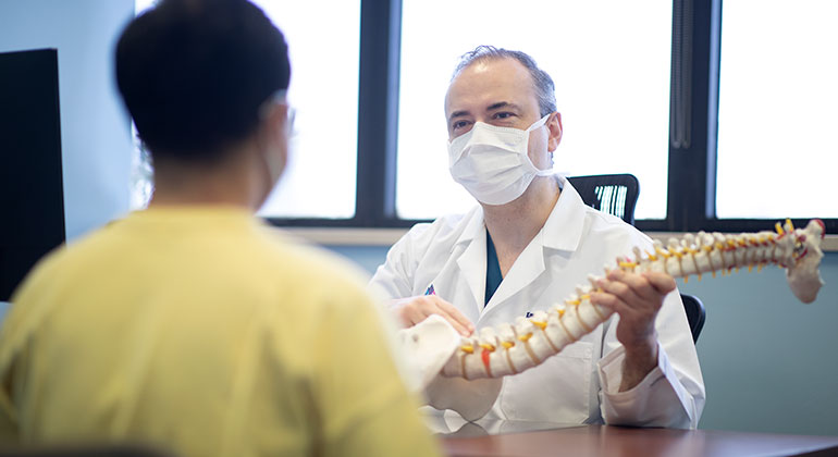
The Department of Neurosurgery at Mount Sinai Health System is the largest program in New York City. Our faculty members have plenty of experience with a wide range of complex cases and are all experts in one or more areas of neurosurgery.
We use a multidisciplinary approach to provide a complete range of diagnostic, treatment, and rehabilitation services for patients with general neurological disorders affecting the brain and spine, including their supportive structures and vascular supply. We use both minimally invasive and traditional open neurosurgical techniques. Our neurosurgeons are also trained and equipped to handle emergency procedures.
General Neurosurgery Conditions
The most common conditions that we treat are: meningiomas, brain tumors, pituitary tumors, chiari malformations, and trigeminal neuralgia.
We treat these various conditions with a variety of treatments including cerebrospinal diversion procedures, disc replacement procedures, minimally invasive spine surgeries, intrathecal pumps (for pain and spasticity), microdiscectomies and spinal fusions, spinal cord stimulators, and vagal nerve stimulators.
Clinical Diagnostic Procedures
Accurate and efficient diagnoses are a vital part of the services we perform. Some of the most common diagnostic procedures are:
Biopsies allow our pathologists to examine a small sample of tissue or cells for any abnormalities. Our neurosurgeons use biopsies to rule out cancer or to specify a type and level of cancer. Biopsies also help us troubleshoot causes of infection, swelling, or growth.
There are various types of biopsies your doctor can order. Some are simple outpatient procedures, while others require general anesthesia and a short hospital stay. Some of the most common types of biopsies are:
- Needle biopsy (fine needle aspiration) involves removing cells using a thin needle.
- Endoscopic biopsy involvesusingalong, thin tube with a camera to navigate to the desired area, and a small tool to collect the sample.
- Excisional biopsy iswherewe remove a relatively largeportion of the mass to get a larger sample.
Computerized tomography (CT) scans are a type of X-ray that uses computers to create images of the inside of your head. We can do CT scans with or without contrast (a chemical that we insert into your vein to give us clear images of certain disorders). A CT scan takes about five minutes and causes little to no pain or discomfort. CT scans can also help us create three-dimensional simulations for surgical planning and medical modeling.
Magnetic resonance imaging (MRI) scans help us diagnose brain injuries and conditions and check the effectiveness of medicines or other treatments. We can use MRIs with contrast for a more detailed image. The test takes between 40 and 90 minutes and is generally painless. We have open MRI machines for patients who are nervous about closed spaces. The MRI can be used to create 3D simulations for surgical planning and medical modeling.
Electrophysiology studies allow us to locate any abnormal electrical signals in your brain and treat the specific areas that are affected by the injured or diseased tissue.
Electroencephalograms (EEG) use sensors that we place on your head to monitor your brain’s electrical activity. EEGs are particularly helpful in diagnosing and determining the side effects of seizures, brain trauma, coma, and brain infections. This hour-long test is painless and you remain awake the entire time.
Angiograms are X-ray exams that use a dye injected into the blood vessels to make the veins and arteries more visible. Angiograms help our surgeons identify disorders of the blood vessels and vascular system. To do this procedure, which usually takes about an hour, we insert a needle into your artery at the groin or arm, then insert a catheter.
Myelography is an imaging test that produces a detailed picture of your spinal cord, nerve roots, and spinal column. We call this picture a myelogram. The test helps us diagnose spinal tumors, herniated disks, and narrowing of the spinal canal. To perform the test, we inject a small amount of contrast dye in your back, then take images of your back. Occasionally, we follow this test with a CT scan so that we can view the spread of the contrast dye. The procedure usually takes 30 to 60 minutes and you can typically return home an hour after the test.
Meet our Faculty
Konstantinos Margetis, MD, PhD
Assistant Professor, Department of Neurosurgery
Fedor E. Panov, MD
Assistant Professor, Department of Neurosurgery
Chanland (Chan) Roonprapunt, MD, PhD
Assistant Professor, Department of Neurosurgery
Raymund L. Yong, MD
Assistant Professor, Departments of Neurosurgery and Oncological Sciences