Transcranial Doppler Ultrasound
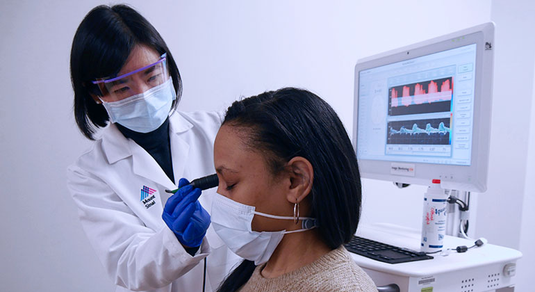
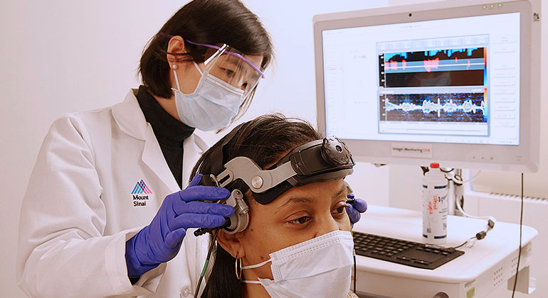
A transcranial doppler (TCD) ultrasound is a safe, non-invasive medical imaging tool that utilizes soundwaves to assess blood flow of the major cerebral arteries within your brain. Using a small device (transducer), sound waves are sent out and then bounced back by the flowing blood, which are turned into graphs or color pictures to assess speed and other features of the blood flow. The features of the blood flow can help to estimate if you have narrowing of the blood vessels and the degree of the narrowing. A TCD can also capture the signal of small blood clots traveling through your blood stream, and the change of blood flow when holding your breath. All this information can help to understand the risk of having a stroke.
Performed in Mount Sinai Neurology’s Neurovascular Laboratory, experienced vascular neurologists will perform the transcranial doppler ultrasound in person, or work with technicians closely, to perform the tests and interpret the results in the setting of clinical context (including neurological symptoms and exam, brain imaging findings), and communicate with your physician who ordered the tests. A full neurovascular work-up, including a Carotid Artery Duplex Ultrasound, may also be performed.
The transcranial doppler may be ordered by your physician to evaluate the blood flow in your brain in certain conditions, including:
- Narrowing of brain blood vessels
- Transient ischemic attack
- Ischemic stroke
- Sickle cell anemia
- Subarachnoid hemorrhage
Transcranial doppler may also be used to assess if there is a small opening between the two chambers of the heart, a condition called Patent Foramen Ovale (PFO).
During Your Exam
With no special preparation needed, the transcranial doppler ultrasound is painless and harmless as sound waves produce the images inside your brain. The exam will take approximately between 30 to 60 minutes.
- After applying a tiny amount of gel, a small device (transducer) will be placed at back of your neck, underneath your jaw, in front of your ears, or over your eyelids to assess the blood flow (the gel will not harm your skin or stain your clothing)
- Though you’ll feel no pain, you may experience mild pressure as the transducer is pressed against your skin
- You will hear a "whooshing" sound, which is normal
- Keep your head still and avoid talking during the exam
- Depending on the purpose of the study, a head frame may be placed around the head to hold the transducers in order to monitor the blood flow continuously for 10- 30 minutes
- If the test is to evaluate PFO, sterile saline filled with tiny bubbles will be injected into your arm vein
Results
After the exam is complete, a vascular neurologist trained specifically in the evaluation and management of cerebrovascular disorders will review the results and determine how the blood flow is, and determine how open, narrow, or blocked your cerebral arteries are.
Normal results
If your vascular neurologist accesses that your results are normal, it means that there is no issue with the blood flow and brain vessels within the brain. Depending on the reason for your exam, you may be requested to come back for additional follow-up tests.
Abnormal results
If your vascular neurologist accesses that your results are abnormal, it means that there are some issues with the blood flow and brain vessels within your brain, including narrowing and blockage, and you may be at a higher risk for stroke.
Conditions that can be detected:
- Narrowing (stenosis) or blockage of brain arteries
- Cerebral microemboli (small blood clots)
- Vasospasm (transient constriction of blood vessel(s) due to a subarachnoid hemorrhage or other conditions)
Follow-Up
Depending on the results of the transcranial doppler ultrasound and the intensity of the narrowing or blockage, your physician may recommend any of the following:
- New medications
- Medications to prevent/slow down the vessel narrowing
- Lifestyle Changes
- Healthy diet and exercise to prevent hardening of the arteries and plaque buildup
- Performing additional tests - CT angiography, and magnetic resonance angiography (MRA), cerebral angiography
- Cerebrovascular Surgery - Requiring follow-up testing and repeating of the ultrasound in the future
A diagnostic ultrasound, also called a sonography, is a safe, non-invasive medical imaging tool that produces images of structures within the body. Using soundwaves to bounce off the blood vessels in your neck, the carotid artery duplex ultrasound is designed to specifically create images and assess vascular blockages or narrowing in the carotid arteries and its branches. The more narrowed your artery is, the higher you’re at risk for a stroke.
Carotid arteries, one located on the right side of the neck and other on the left, are critical blood vessels which supply blood to the head and brain. The arteries in the neck branch; the internal carotid arteries supply blood to the brain while the external carotid artery supplies blood to the face and neck.
Performed in Mount Sinai Neurology’s Neurovascular Laboratory, experienced stroke neurologists will perform the ultrasound in person, and/or work with technicians closely, to perform tests and interpret the carotid duplex in the setting of clinical context (including neurological symptoms and exam, brain imaging findings), and communicate with your physician who ordered the tests. A full neurovascular work-up, including a Transcranial Doppler Ultrasound, may also be performed.
The carotid artery duplex ultrasound may be ordered by your physician to check the blood flow and blood vessels in your carotid artery to either diagnose or have a follow-up monitoring, because you may have:
- Presented with symptoms including vision disturbance, arm or leg weakness, speech abnormalities, dizziness, balance issues, or memory loss
- Abnormal sound called bruit in the neck region during the physical exam
- Had a previous stroke or transient ischemic attack (TIA)
- Previously detected a narrowing in your carotid artery
- Had surgery on your carotid artery
During Your Exam
With no special preparation needed, the carotid artery duplex ultrasound is painless and harmless as sound waves produce the images of the blood vessels inside of your neck. The exam will take approximately between 15 to 30 minutes.
- We recommend not wearing necklace, and wearing looser clothing, especially around the neck
- Warm gel is applied to the neck area
- Though you’ll feel no pain, you may experience mild pressure as the transducer is pressed against your neck
- You will hear a "whooshing" sound, which is normal
- Keep your head still and avoid talking during the exam
Results
After the exam is complete, a vascular neurologist trained specifically in the evaluation and management of cerebrovascular disorders will review the results and determine how open, narrow, or blocked your carotid arteries are.
Normal Results
If your vascular neurologist accesses that your results are normal, it means that there is no issue with the carotid arteries or their branches. Depending on the reason for your exam, you may be requested to come back for additional follow-up tests.
Abnormal Results
If your vascular neurologist accesses that your results are abnormal, it means that there are some issues with the carotid arteries or their branches, including narrowing and blockage, and you may be at a higher risk for stroke.
Conditions that can be detected:
- Atherosclerosis (plaque buildup in the arteries)
- Thrombosis (blood clotting)
- Dissection (tear of the vessel wall)
- Other cause of narrowing (stenosis) or blockage in the carotid arteries
Follow-Up
Depending on the results of the carotid artery duplex ultrasound and the intensity of the narrowing or blockage, your physician may recommend any of the following:
- Medications to prevent/slow down the vessel narrowing
- Lifestyle Change
- Healthy diet and exercise to prevent hardening of the arteries and plaque buildup
- Performing of Additional Tests - CT angiography, and magnetic resonance angiography (MRA), cerebral angiography
- Cerebrovascular Surgery - requiring follow-up testing and repeating of the ultrasound in the future