Renal venogram
Venogram - renal; Venography; Venogram - kidney
A renal venogram is a test to look at the veins in the kidney. It uses x-rays and a special dye (called contrast).
X-rays are a form of electromagnetic radiation like light, but of higher energy, so they can move through the body to form an image. Structures that are dense (such as bone) will appear white and air will be black. Other structures will be shades of gray.
Veins are not normally seen in an x-ray. That is why the special dye is needed. The dye highlights the veins so they show up better on x-rays.
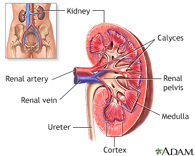
The kidneys are responsible for removing wastes from the body, regulating electrolyte balance and blood pressure, and the stimulation of red blood cell production.
A renal venogram is a method used to examine the veins of the kidneys, using a contrast material and x-rays.
How the Test is Performed
This test is done in a health care facility with special equipment. You will lie on an x-ray table. Local anesthetic is used to numb the area where the dye is injected. You may ask for a calming medicine (sedative) if you are anxious about the test.
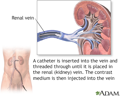
The health care provider places a needle into a vein, most often in the groin, but occasionally in the neck. Next, a flexible tube, called a catheter (which is the width of the tip of a pen), is inserted into the groin and moved through the vein until it reaches the vein in the kidney. A blood sample may be taken from each kidney. The contrast dye flows through this tube. X-rays are taken as the dye moves through the kidney veins.
This procedure is monitored by fluoroscopy, a type of x-ray that creates images on a TV screen.
Once the images are taken, the catheter is removed and a bandage is placed over the wound.
How to Prepare for the Test
You will be told to avoid food and drinks for about 6 hours before the test. Your provider may tell you to stop taking aspirin or other blood thinners before the test. DO NOT stop taking any medicine without first talking to your provider.
You will be asked to wear hospital clothing and to sign a consent form for the procedure. You will need to remove any jewelry from the area that is being studied.
Tell the provider if you:
- Are pregnant
- Have allergies to any medicine, contrast dye, or iodine
- Have a history of bleeding problems
How the Test will Feel
You will lie flat on the x-ray table. There is often a cushion, but it is not as comfortable as a bed. You may feel a sting when the local anesthesia medicine is given. You will not feel the dye. You may feel some pressure and discomfort as the catheter is positioned. You may feel symptoms, such as flushing, when the dye is injected.
There may be mild tenderness and bruising at the site where the catheter was placed.
Why the Test is Performed
This test is not done very often anymore except in the treatment of varicose veins of the testicles or ovaries. Otherwise, it has largely been replaced by CT scan and MRI. In the past, the test was used to measure levels of kidney hormones.
Rarely, the test may be used to detect blood clots, tumors, and vein problems. Its most common use today is as part of an exam to treat varicose veins of the testicles or ovaries.
Normal Results
There should not be any clots or tumors in the kidney vein. The dye should flow quickly through the vein and not back up to the testes or ovaries.
What Abnormal Results Mean
Abnormal results may be due to:
Risks
Risks from this test may include:
- Allergic reaction to the contrast dye
- Bleeding
- Blood clots
- Injury to a vein
There is low-level radiation exposure. However, most experts feel that the risk of most x-rays is smaller than other risks we take every day. Pregnant women and children are more sensitive to the risks of the x-ray.
References
Bechara CF. Venography. In: Sidawy AN, Perler BA, eds. Rutherford's Vascular Surgery and Endovascular Therapy. 10th ed. Philadelphia, PA: Elsevier; 2023:chap 28.
Perico N, Remuzzi A, Remuzzi G. Pathophysiology of proteinuria. In: Yu ASL, Chertow GM, Luyckx VA, Marsden PA, Skorecki K, Taal MW, eds. Brenner and Rector's The Kidney. 11th ed. Philadelphia, PA: Elsevier; 2020:chap 30.
Wymer DTG, Wymer DC. Radiologic and nuclear imaging in nephrology. In: Johnson RJ, Floege J, Tonelli M, eds. Comprehensive Clinical Nephrology. 7th ed. Philadelphia, PA: Elsevier; 2024:chap 6.
Version Info
Last reviewed on: 1/29/2024
Reviewed by: Jason Levy, MD, FSIR, Northside Radiology Associates, Atlanta, GA. Also reviewed by David C. Dugdale, MD, Medical Director, Brenda Conaway, Editorial Director, and the A.D.A.M. Editorial team.
