Cellular Atlas of a Human Brain Language Area Is Constructed by a Team of International Scientists
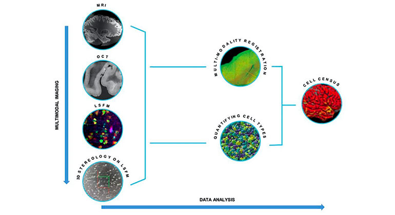
Imaging and analysis overview. From top to bottom, multimodal imaging. MRI, OCT, LSFM, and 3D stereology on LSFM images are performed on the same sample. Left to right, data analysis. Registration between MRI, OCT, LSFM and 3D stereology data is performed to align all the datasets back to the MRI coordinate system. Cell counting on LSFM images with digital stereology reliable quantification of cell types. Multimodal registration between the images and the counting is needed to finally obtain the cell census of the neurons in an MRI-based atlas coordinate system.
A global team of researchers that includes Mount Sinai scientists has created a comprehensive cellular atlas of a part of the cerebral cortex of the human brain at the single-cell level, opening the door to characterizing additional areas of the brain and potentially identifying markers for disorders as well as innovative new treatments.
As described in Science Advances, the scientists used advanced imaging and analysis techniques to describe the cellular architecture of a region of the frontal lobe of the brain known as Broca’s area, giving them a revealing window on how those cells are arranged and their associated functions. Broca’s area is known as the motor area responsible for speech production, with linkage to language processing and comprehension.
The paper is included in a package of 21 research studies across Science, Science Advances and Science Translational Medicine that detail research conducted as part of the National Institutes of Health’s BRAIN Initiative Cell Census Network (BICCN), a program launched in 2017 to create an atlas of the human and non-human primate brain at the cell-type level in unprecedented detail.
“The human brain is a complex organ spanning an astonishing range of spatial scales, and to understand its properties and functionality it’s essential to construct a detailed census of its many classes of neurons and visualize their distribution across the whole brain volume,” says Patrick R. Hof, MD, Dorothy and Irving Regenstreif Research Professor of Neuroscience (Neurobiology) at the Icahn School of Medicine at Mount Sinai, and senior author of the study.
“Our multi-institutional team developed a novel way to document and quantify the cellular organization of neurons at the micrometer level, and we believe this approach will allow us to gain fundamental information about the cellular architecture of various areas of the brain,” Dr. Hof says. “These could include subcortical domains and even entire hemispheres.”
In addition to the Nash Family Department of Neuroscience and The Friedman Brain Institute at Icahn Mount Sinai, institutions that were part of the collaborative team included the Department of Radiology, Athinoula A. Martinos Center for Biomedical Imaging at Massachusetts General Hospital; the Department of Biomedical Engineering at Boston University; the European Laboratory of Non-Linear Spectroscopy and the Department of Biology at the University of Florence, Italy; and the Department of Medical Physics and Biomedical Engineering at University College, London.
The work focused on cellular diversity of the cerebral cortex and, more specifically, Broca’s area. The creation of a detailed reference map of this complex sphere required the research team to build 3D models of the human brain with cellular resolution through the use of magnetic resonance imaging (MRI).
“While advances in technology have made it possible to obtain a comprehensive cell census in animal models, no imaging modalities existed to directly visualize the microscopic features of the whole human brain without significant distortion,” explained Dr. Hof, who directs the Kastor Neurobiology of Aging Laboratories at Mount Sinai. “With our colleagues, we overcame these limitations by creating a multimodal imaging infrastructure to bridge the resolution gap between macroscopic and microscopic techniques.”
Essential to that construct was the application by the Boston University team of optical coherence tomography (OCT), a non-invasive optical imaging technique that uses backscattered light in the near-infrared spectral range to provide high-resolution, cross-sectional images and volumetric reconstruction of brain tissue up to several hundred micrometers in depth. “Using OCT as an intermediate modality allowed us to successfully collect the quantitative cell census of different neuronal types in the Broca’s area within the whole-hemisphere MRI reference framework of a postmortem human brain,” Dr. Hof notes.
Having demonstrated that a cellular atlas of one identifiable cortical region could be achieved, the global research team is now prepared to apply its findings to other unexplored realms of the brain. Their study suggests that comparable methods could be used to study the brain’s connectivity, thereby resolving not only cell types but pathways and networks at the microscopic level. Moreover, future studies might analyze brain tissue from not just neurotypical donors, but patients who experienced different neuropsychiatric diseases, providing information about neuropathologic alterations of cellular organization in brain disorders.
“We’ve developed a new imaging pipeline for characterizing the brain cytoarchitecture, and plan to leverage that knowledge to construct even more comprehensive atlases, including new markers and analyses of connectivity,” Dr. Hof emphasizes. “Moreover, this approach could be used by other laboratories to scale up their own analyses and increase our knowledge about the brain.”
Research reported in this press release was supported by the NIH BRAIN Initiative under award number: U01 MH117023. This publication was supported by and coordinated through the BICCN (biccn.org). This publication is part of the Human Cell Atlas (www.humancellatlas.org/publications/).
About the Mount Sinai Health System
Mount Sinai Health System is one of the largest academic medical systems in the New York metro area, with more than 43,000 employees working across eight hospitals, over 400 outpatient practices, nearly 300 labs, a school of nursing, and a leading school of medicine and graduate education. Mount Sinai advances health for all people, everywhere, by taking on the most complex health care challenges of our time — discovering and applying new scientific learning and knowledge; developing safer, more effective treatments; educating the next generation of medical leaders and innovators; and supporting local communities by delivering high-quality care to all who need it.
Through the integration of its hospitals, labs, and schools, Mount Sinai offers comprehensive health care solutions from birth through geriatrics, leveraging innovative approaches such as artificial intelligence and informatics while keeping patients’ medical and emotional needs at the center of all treatment. The Health System includes approximately 7,300 primary and specialty care physicians; 13 joint-venture outpatient surgery centers throughout the five boroughs of New York City, Westchester, Long Island, and Florida; and more than 30 affiliated community health centers. We are consistently ranked by U.S. News & World Report's Best Hospitals, receiving high "Honor Roll" status, and are highly ranked: No. 1 in Geriatrics and top 20 in Cardiology/Heart Surgery, Diabetes/Endocrinology, Gastroenterology/GI Surgery, Neurology/Neurosurgery, Orthopedics, Pulmonology/Lung Surgery, Rehabilitation, and Urology. New York Eye and Ear Infirmary of Mount Sinai is ranked No. 12 in Ophthalmology. U.S. News & World Report’s “Best Children’s Hospitals” ranks Mount Sinai Kravis Children's Hospital among the country’s best in several pediatric specialties.
For more information, visit https://www.mountsinai.org or find Mount Sinai on Facebook, Twitter and YouTube.