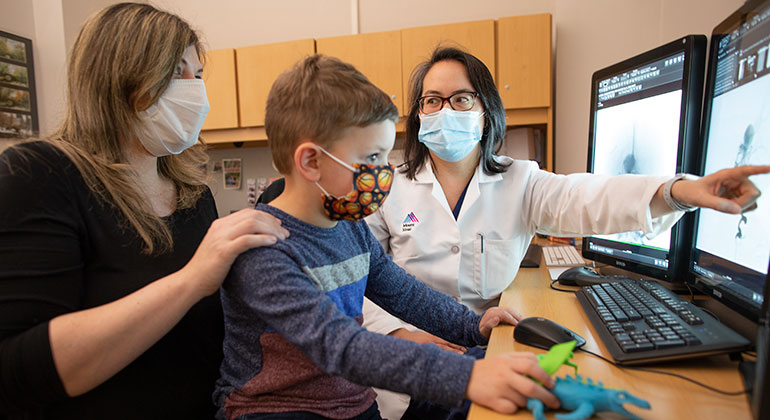
At the Cerebrovascular Center at Mount Sinai, we are leaders in the field of cerebrovascular surgery. We offer the least invasive treatments appropriate for your condition. We use endovascular therapy, radiosurgery, open microsurgery, or a combination of these techniques. These approaches reduce risk of complications, and offer a shorter, less painful recovery period.
We regularly hold multidisciplinary conferences to determine the complexity and severity of your disorder and decide on the best treatment plan. We use the following approaches:
Endovascular Procedures
Minimally invasive endovascular procedures allow our team of specialists to guide catheters, microcatheters, and other devices through your blood vessels to address pressing health concerns. Typically, we enter through the femoral artery in the groin or radial artery in the wrist, using fluoroscopic (X-ray) guidance. We use several types of endovascular procedures:
Angiograms help us to visualize blood vessels by X-ray. To perform an angiogram, we use a local anesthesia in the upper part of your thigh. Then we insert a catheter into your body through a blood vessel in that area. We administer intravenous sedation to keep you comfortable and make sure you do not move while we take the X-rays.
We use X-ray guidance to carefully thread the catheter through the blood vessels until it reaches the area of interest. Once the catheter tip is in position, we inject the contrast material through the catheter to the spot while taking a rapid series of x-ray images. The dye highlights the blood vessels we need to see. While you probably will not feel the catheter, you may have a sensation of warmth when we inject the contrast dye.
Once we finish the angiogram, we remove the catheter. You will probably stay at the hospital a few hours so we can observe you. You will likely go home later that day.
Angioplasty, also called a balloon angioplasty, helps us reopen clogged blood vessels of the arteries of the neck and brain. The carotid artery supplies blood from the heart to the brain; unblocking a narrowed carotid artery helps prevent strokes.
During this minimally invasive procedure, we make a tiny incision in the skin of the groin, then insert a catheter with an angioplasty balloon at the tip. We thread the catheter through the femoral artery, a large blood vessel in the leg, until we get it to the point where the artery has become narrow. We use imaging techniques such as X-rays to guide us. Once the catheter is properly positioned, we inflate the angioplasty balloon, which opens the narrowing. Sometimes, we combine this approach with stent technology, a procedure where we insert a small metal mesh in the artery to keep it propped open.
Embolization is when we deliberately close a blood vessel to treat a condition such as cerebral (brain) aneurysms, arteriovenous malformations (or AVMs) of the brain and spine, vein of Galen malformations, vascular malformations, tumors and nosebleeds. Sometimes we only need to do an embolization, but other times we use this technique along with surgery. We can also use embolization to improve quality of life (palliative), such as reducing pain.
We perform embolization by inserting a catheter through a tiny incision in the skin of the groin into the femoral artery, a large blood vessel in the leg. Using fluoroscopy or X-ray guidance, we lead the catheter to the site of the abnormality through your blood vessels, until the tip of the catheter is at the problem site. Then we inject an embolizing agent through the catheter, to seal the blood vessel. We use a variety of embolic agents, including small plastic particles, glue, metal coils, foam, and/or a balloon, depending on the situation.
Infusion therapy, also called chemotherapy helps treat cancerous tumors. We administer it either intravenously or orally. The medication travels through the bloodstream throughout the body. Both approaches have two major (and interrelated) drawbacks. First, the medicine becomes diluted in the bloodstream, so the tumor receives a smaller dose of cancer-fighting medicine than what we administer. Second, most chemotherapy therapies produce a variety of side effects. Typically, if we boost increase the dose so the tumor receives more medication, the side effects increase as well.
Infusion therapy allows us to deliver chemotherapy directly to the site of the tumor, which increases the amount of medicine directed at the tumor and decreases the side effects.
To perform infusion therapy, we use endovascular techniques to place a catheter is placed into the artery that feeds the tumor and inject the chemotherapeutic medication directly into that blood vessel.
Sclerotherapy is when we inject a substance to scar vascular malformations and hemangiomas. By scarring the highly vascular malformation, we reduce the blood supply and flow velocity to the problem site, which makes it shrink. We perform this procedure using ultrasound guidance and fluoroscopy or real-time X-ray monitoring. We use a variety of sclerosing agents, depending on the individual situation.
Endovascular Laser Therapy Ablation is an option for large venous malformations, especially when schlerotherapy is not a viable choice. By also using endovenous laser therapy ablation, we can speed up the response and ease the recovery process. Extensive vascular malformations often require a series of these procedures, and we may also perform additional treatment a few years later.
It may take months to fully heal from the combination of endovascular laser therapy and sclerotherapy. You will likely experience swelling and bruising, which will start to go down over the first day or two. The swelling will not go completely away until the blood within the damaged site is absorbed, which may take two to three months. If the skin is involved in your vascular malformation, you may develop a blister or sore with some discoloration. We can provide pain medication during the healing process.
Radiosurgery
Radiosurgery is appropriate for vascular malformations in the deepest locations in the brain. We direct multiple low-power radiation beams so that they overlap right at the vascular malformation. This approach treats the malformation while protecting the surrounding normal tissue.
Open Procedures
Minimally invasive open surgical techniques use incisions that are 70 percent smaller than with traditional techniques, resulting in less tissue injury, better cosmetic results, and fast recovery.
One approach to open procedures is the use of stenting. Stenting allows us to unblock the carotid artery in the neck, which is a major supplier of blood to the brain. Unblocking this artery with a carotid artery stent helps prevent strokes.
To use stents, we insert a catheter through a tiny incision in the skin of the groin into the femoral artery, a large blood vessel in the leg. Guided by imaging techniques such as X-rays, we thread the catheter through the body's circulatory system until the tip of the catheter is just below the narrowing of the carotid artery or arteries of the brain. We place a wire mesh tube, called a stent, in the blocked area. This approach is becoming more and more common.
Vagus Nerve Stimulation for Stroke Rehabilitation
After experiencing a stroke, many survivors continue to suffer from persistent impaired upper limb function and weakness. Vagus Nerve Stimulation (VNS) electrical pulses release neuromodulators in the brain that create or strengthen neural connections to enhance the relevance of physical therapy and improve upper limb function. “Pairing VNS with rehabilitation helps stroke survivors regain their independence and improve their quality of life, months or even years after suffering from a stroke,” says cerebrovascular neurosurgeon Christopher Kellner, MD Co-Founder of the Enhanced Stroke Recovery Program. More information about paired vagus nerve stimulation for stroke rehabilitation.
Frequently Asked Questions
What is endovascular surgery?
Endovascular surgery, a type of minimally invasive surgery, requires only a small incision in the groin to gain access to a problematic location in the body. It eliminates the need for traditional surgery. We sometimes call endovascular procedures "image-guided surgery" since the doctor can see where the catheter is going and what is happening at all times. Major advances in imaging equipment have led to great improvements and new possibilities in the field of endovascular surgery.
How do I find out if I am a candidate for endovascular surgery?
Our doctors work closely with your established doctor to learn about the history and background of your condition. Our nursing staff will coordinate this process when they speak with you. They will either ask permission to contact your present doctor or set up a clinic appointment for you to see one of our doctors. At that appointment, our doctor will determine what additional tests they need and explain your treatment options. If your condition is unusually complex, we may review your case at our weekly multidisciplinary physician meeting.
What can I expect before the procedure?
If our doctors can help you, we will conduct a series of pre-procedure exams and laboratory tests. On the day of the procedure, you will register with the hospital and our staff will direct you to the appropriate area.
How long will I be hospitalized?
Most people stay in the hospital for one to five days, though this can vary greatly depending on your condition and general health.
What can I expect after the procedure?
After you leave the hospital, we will monitor you closely for the next year, starting at four weeks after the procedure. We will use magnetic resonance imaging and X-rays periodically.