Gallstones
Cholelithiasis; Gallbladder attack; Biliary colic; Gallstone attack; Biliary calculus: gallstones chenodeoxycholic acids (CDCA); Ursodeoxycholic acid (UDCA, ursodiol); Endoscopic retrograde cholangiopancreatography (ERCP) - gallstones
Gallstones are hard deposits that form inside the gallbladder. These may be as small as a grain of sand or as large as a golf ball.
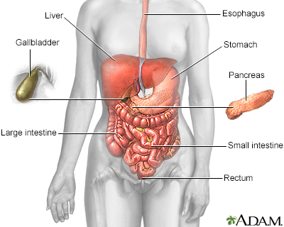
The esophagus, stomach, large and small intestine, aided by the liver, gallbladder and pancreas convert the nutritive components of food into energy and break down the non-nutritive components into waste to be excreted.
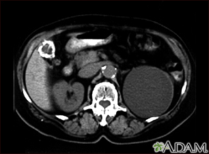
A CT scan of the upper abdomen showing a fist-sized cyst of the left kidney and gallstones (the kidney cyst was found by chance; there were no symptoms).
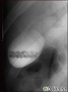
A cholecystogram in a patient with gallstones.
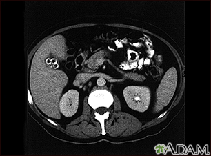
Cholecystolithiasis. CT scan of the upper abdomen showing multiple gallstones.
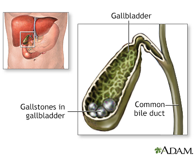
Normally a balance of bile salts, lecithin and cholesterol keep gallstones from forming. If there are abnormally high levels of bile salts or, more commonly, cholesterol, stones can form. Symptoms usually occur when the stones block one of the biliary ducts or gallstones may be discovered upon routine x-ray or abdominal CT study.
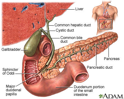
The gallbladder is a muscular sac located under the liver. It stores and concentrates the bile produced in the liver that is not immediately needed for digestion. Bile is released from the gallbladder into the small intestine in response to food. The pancreatic duct joins the common bile duct at the small intestine adding enzymes to aid in digestion.
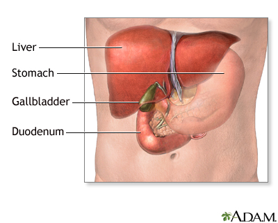
The gallbladder is located in the abdomen on the underside of the liver. The gallbladder stores bile (digestive fluid) from the liver.
Causes
The cause of gallstones varies. There are two main types of gallstones:
- Stones made of cholesterol -- This is the most common type. Cholesterol gallstones are not related to cholesterol level in the blood. In most cases, they are not visible on CT scans but are visible on a sonogram (ultrasound) of the abdomen.
- Stones made of bilirubin -- These are called pigment stones. They occur when red blood cells are destroyed and too much bilirubin is in the bile.
Gallstones are more common in:
- Female sex
- Native Americans and people of Hispanic descent
- People over age 40
- People who are overweight
- People with family history of gallstones
The following factors also make you more likely to develop gallstones:
- Bone marrow or solid organ transplant
- Diabetes
- Failure of the gallbladder to empty bile properly (this is more likely to happen during pregnancy)
- Liver cirrhosis and biliary tract infections (pigmented stones)
- Medical conditions that cause too many red blood cells to be destroyed
- Rapid weight loss from eating a very low-calorie diet, or after weight loss surgery
- Receiving nutrition through a vein for a long period of time (intravenous feedings)
- Taking birth control pills
Symptoms
Many people with gallstones do not have any symptoms. These are often found during a routine x-ray, abdominal surgery, or other medical procedure.
However, if a large stone blocks a tube or duct that drains the gallbladder, you may have a cramping pain in the middle to right upper abdomen. This is known as biliary colic. The pain goes away if the stone passes into the first part of the small intestine.
Symptoms that may occur include:
- Pain in the right upper or middle upper abdomen for at least 30 minutes. The pain may be constant or cramping. It can feel sharp or dull.
- Fever.
- Yellowing of skin and whites of the eyes (jaundice).
Other symptoms may include:
- Clay-colored stools
- Nausea and vomiting
Stones are great for tossing into a stream or using to line your flowerbeds. But they're not so great when they're trapped inside your gallbladder. If you've got pain in the upper part of your belly, a fever, or yellow skin, they could be signs that you've got gallstones. The stones that form in your gallbladder aren't made of rock. Usually, they're made of cholesterol, a type of fat in your blood. Or, they could be made from a substance called bilirubin, which is processed in your liver. Gallstones can be as small as a grain of sand, or as big as a golf ball. You're more likely to get gallstones if you're over age 40, or if you have a chronic condition like diabetes, anemia, or cirrhosis of the liver. People who've had weight-loss surgery, or who went on a crash diet and lost weight very quickly can also develop gallstones. Gallstones are more common in women than in men, and they may run in families. So, how do you know if you have gallstones? Well, you might not realize it, because often gallstones don't cause any pain. They're often found accidentally during an x-ray or surgery to treat another condition. If the gallstone is very large, though, it may get stuck in one of the tubes, called ducts, which connect to the gallbladder. Then you'll probably feel a sharp or cramping pain in the upper right or middle part of your abdomen. You may also have a fever and feel sick to your stomach. Your doctor can do an ultrasound, or other scan of the gallbladder area to find out if gallstones are causing your pain. You may also have blood tests to check your liver function and to see if your bilirubin levels are too high. You may not need to treat gallstones, unless they're causing symptoms. If that's the case, your doctor will recommend treatment which is usually surgery to remove them. Often, this is done with a laparoscopic procedure that removes the gallstone through very small cuts or incisions. Normally, you can go home the same day as your surgery, or the next day. Most people don't need to have their whole gallbladder removed, unless they have complications, like a blocked duct. There is also medicine that can dissolve cholesterol gallstones, but it isn't that effective because it can take two years or more to work, and often the stones form again after you're done taking it. You can't really prevent gallstones, except by avoiding rapid weight loss or health conditions that can cause gallstones such as obesity, diabetes, or cirrhosis. But if you do get them, gallstones are pretty easy to treat. Most people don't have any symptoms or complications from them, and those who do have symptoms usually recover completely and don't get gallstones again after their surgery. It's important that you call your doctor if you are having abdominal pain, yellow skin or eyes, so you can find out for sure whether you have gallstones, and get them treated.
Exams and Tests
Tests used to detect gallstones or gallbladder inflammation include:
- Ultrasound, abdomen
- CT scan, abdomen
- Endoscopic retrograde cholangiopancreatography (ERCP)
- Gallbladder radionuclide scan
- Endoscopic ultrasound
- Magnetic resonance cholangiopancreatography (MRCP)
- Percutaneous transhepatic cholangiogram (PTCA)
Your health care provider may order the following blood tests:
- Bilirubin
- Liver function tests
- Complete blood count
- Pancreatic enzyme (amylase or lipase)
Treatment
SURGERY
Most of the time, surgery is not needed unless symptoms begin. However, people planning weight loss surgery may need to have gallstones removed before undergoing the procedure. In general, people who have symptoms will need surgery right away or soon after the stone is found.
- A technique called laparoscopic cholecystectomy is most commonly used. This procedure uses small surgical incisions, which allow for a faster recovery. A patient can often go home from the hospital within 1 day of surgery.
- In the past, open cholecystectomy (gallbladder removal) was most often done. However, this technique is less common now.
ERCP and a procedure called a sphincterotomy may be done to find or treat gallstones in the common bile duct.
MEDICINES
Medicines may be given in pill form to dissolve cholesterol gallstones. However, these drugs may take 2 years or longer to work, and the stones may return after treatment ends.
Rarely, chemicals are passed into the gallbladder through a catheter. The chemical rapidly dissolves cholesterol stones. This treatment is hard to perform, so it is not done very often. The chemicals used can be toxic, and the gallstones may return.
LITHOTRIPSY
Shock wave lithotripsy (ESWL) of the gallbladder has also been used for people who cannot have surgery. This treatment is not used as often as it once was because gallstones often come back.
Outlook (Prognosis)
You may need to be on a liquid diet or take other steps to give your gallbladder a rest after you are treated. Your provider will give you instructions when you leave the hospital.
The chance of symptoms or complications from gallstone surgery is low. Nearly all people who have their gallbladder taken out by surgery do not have their symptoms return.
Possible Complications
Blockage by gallstones may cause swelling or infection in the:
- Gallbladder (cholecystitis)
- Tube that carries bile from the liver to the gallbladder and intestines (cholangitis)
- Pancreas (pancreatitis)
When to Contact a Medical Professional
Contact your provider if you have:
- Pain in the upper part of your abdomen
- Yellowing of the skin or whites of the eyes
Prevention
In most people, gallstones can't be prevented. In people who are obese, avoiding rapid weight loss may help prevent gallstones.
References
Fogel EL, Sherman S. Diseases of the gallbladder and bile ducts. In: Goldman L, Schafer AI, eds. Goldman-Cecil Medicine. 26th ed. Philadelphia, PA: Elsevier; 2020:chap 146.
Radkani P, Hawksworth J, Fishbein T. Biliary system. In: Townsend CM Jr, Beauchamp RD, Evers BM, Mattox KL, eds. Sabiston Textbook of Surgery. 21st ed. St Louis, MO: Elsevier; 2022:chap 55.
Wang D Q-H, Afdhal NH. Gallstone disease. In: Feldman M, Friedman LS, Brandt LJ, eds. Sleisenger and Fordtran's Gastrointestinal and Liver Disease: Pathophysiology/Diagnosis/Management. 11th ed. Philadelphia, PA: Elsevier; 2021:chap 65.
Version Info
Last reviewed on: 5/2/2023
Reviewed by: Michael M. Phillips, MD, Emeritus Professor of Medicine, The George Washington University School of Medicine, Washington, DC. Also reviewed by David C. Dugdale, MD, Medical Director, Brenda Conaway, Editorial Director, and the A.D.A.M. Editorial team.
