Skull Base Surgery Center
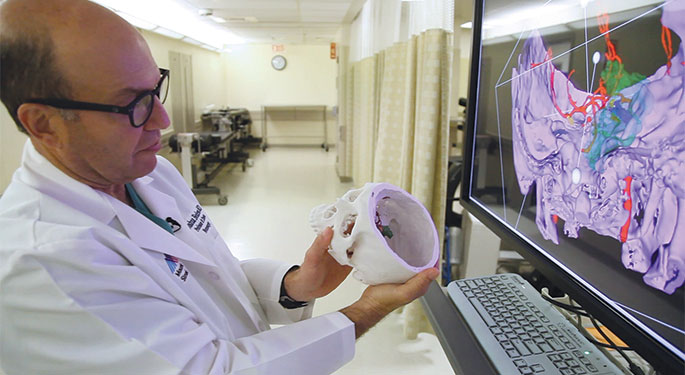
Led by ENT/Head and Neck Surgery Chairman Eric M. Genden, MD, and Neurosurgery Chairman Joshua B. Bederson, MD, the Skull Base Surgery Center at Mount Sinai is one of the few multidisciplinary centers throughout the world dedicated to the evaluation and treatment of benign and malignant tumors of the skull base (floor of the cranium). We have a rich history as a leader in the treatment of these tumors, which continues today through the use of transoral robotic surgery (TORS), transnasal endoscopic brain and pituitary surgery, 3-D navigation planning, intraoperative neurophysiological monitoring (IONM), surgical simulation, and cutting edge research. Our experts craft personalized plans for each patient using the least invasive and safest approach possible. Read more