Brain Tumor Treatments and Procedures
At Mount Sinai, we are pioneers in computer-assisted stereotactic techniques. These approaches allow us to operate on tumors previously thought inoperable. We use techniques such as state-of-the-art navigational microscopes and a computerized navigation system.
When possible, we perform brain mapping to preserve brain function during the surgical removal of brain tumors. Brain mapping reduces surgical risk and improves patient outcome. We perform endoscopic surgery when applicable to remove tumors less invasively and more accurately. This less invasive surgery is well tolerated and promotes speedy recovery. We perform the following types of treatment for brain tumors.
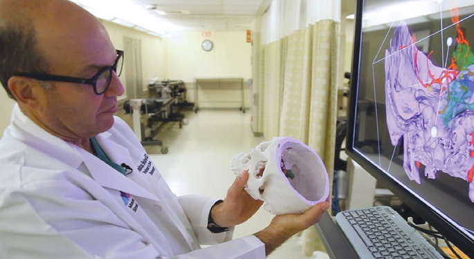
Computer-assisted, image-guided craniotomy helps us gather and store images from your magnetic resonance imaging and computed tomography scans. We can reformat the images so that we can see your lesion three dimensionally, in relation to critical areas of your brain. The technique helps us plan and simulate the surgical procedure beforehand, reach deep-seated or centrally located brain tumors, and employ the safest and least invasive route possible. It also reduces surgical complications, shortens hospital stays and costs, and enhances quality of life. Most important, we can see this information during surgery, scaled to the actual size and location within the surgical field. The scans can guide our actions very accurately, increasing the amount of tumor we can safely remove.
Computer-assisted neurosurgery is appropriate for anybody who need brain surgery and wants a precise and minimally invasive operation. This includes people with brain tumors of all types. Computer-assisted neurosurgery makes it easier to:
-
Use small skin and bone openings
-
Find the lesion quickly
-
Remove the tumor without touching the healthy parts of the brain
-
Plan and simulate the surgery beforehand
-
Know exactly where tumor ends and normal brain begins, which decreases the risk to the surrounding brain tissue
-
Get better results so that you need less rehabilitation and can return to work sooner than with traditional procedures
Awake and asleep brain mapping are advanced neurosurgical procedures to remove brain tumors. During awake brain surgery, you remain alert and responsive at various times while we perform the procedure. This approach is particularly appropriate for brain tumors infiltrating into the parts of the brain responsible for producing and understanding language, which are located in the left frontal and temporal lobes in most right-handed people. These procedures are also effective if we need to search for the areas of the brain responsible for seizures (epilepsy). Having you awake helps us more precisely locate the boundary between the tumor and critical structures near the surface and deep within the brain. There are also many functions, such as movement, hearing, and vision, which we can map or monitor while you are asleep. The more precise we can be, the safer the procedure. Brain mapping usually occurs in conjunction with a computer-assisted imaged-guided craniotomy.
Stereotactic needle biopsy is appropriate when we suspect that there is a brain tumor anywhere in your body, to diagnose the tumor type and guide further care. Biopsies in the brain are complicated, so we routinely use computer-assisted image guidance to direct the procedure.
Stereotactic radiosurgery uses radiation to shrink or destroy malignant tumors. We use it as an alternative to, or in addition to craniotomy or brain biopsy. Stereotactic radiosurgery allows us to deliver precise, concentrated, highly focused radiation directly to the brain tumor while sparing the surrounding tissue. We use low-dose radiation beams to penetrate your brain from different angles. The dosage is low enough to ensure that the healthy brain tissue receives minimal exposure, but significant enough to shrink or kill the tumor at the point where the beams intersect, at the tumor site. We customize the radiation dosage based on the type and size of the tumor. We use advanced technology such as the Novalis Shaped Beam Surgery System. We perform radiosurgery in between one and five sessions, in an outpatient setting.
Radiotherapy is a different procedure than radiosurgery. With radiotherapy, we deliver radiation in 10 to 30 small doses (called fractions) over a two- to six-week period. The radiation targets the region of the tumor as well as the immediate surroundings, which can be advantageous in infiltrative types of brain tumors such as glioblastoma. Radiotherapy typically begins within two to six weeks after surgery and we perform it in the outpatient setting.
Chemotherapy, targeted therapy, and immunotherapy (including vaccines) can be important components of treatment for brain tumors and are aimed at destroying residual cancer cells or keeping them from regrowing. You may require these treatments alone, or in combination with radiation therapy. Typically, these treatments follow diagnostic surgery. We usually provide these treatments in pill form or using an intravenous infusion. In some cases, we use wafers, which we place during surgery. We are involved with clinical trials to test new chemotherapy, targeted therapy, and immunotherapy strategies. If they are appropriate for you and your condition, we will discuss them with you.
Endoscopic Transnasal Resection is a surgical procedure that goes through the nose in a minimally invasive way to remove a tumor or a cyst. Tumors may also be removed without affecting surrounding areas. Most patients can be discharged 2 to 3 days after surgery.
Surgery Instructions
It can be helpful to know what to expect and how to prepare for surgery and recuperation.
Before the procedure
You will meet with your surgeon to discuss the type of tumor you have and what procedure we will perform. We will schedule your surgery give you instructions to make sure you are healthy enough to have the procedure performed safely. You may need to get bloodwork, additional imaging studies, and a check-up from a primary care doctor or specialist if you have a pre-existing condition. We will give you a personalized medical plan to prepare for the procedure.
Day of surgery
We will tell you when to arrive at the hospital and where to go. After you check in, nurses will prepare you for the operating room (OR). You and your family will meet with your surgeon, anesthesiologist, and the rest of the operating room team. The staff will then take you into the OR and show your family members where they can wait. We will let your family know when your procedure is finished.
Recuperation in the hospital
The type of tumor you have and its location in your brain will determine how long you have to stay in the hospital after the procedure, but you can plan on at least one or two days. During this time, our nurses will monitor you closely and you will work with therapists to speed your recovery. We may perform additional testing to monitor your progress. Before you leave, we will give you a discharge plan, which may include transfer to a rehabilitation or skilled nursing facility.
Recuperation at home
Your surgeon will tell you how to take care of your incision at home, what medications to take, what you can and cannot eat, and any precautions you should take. We will schedule a follow-up appointment with your surgeon to discuss your results and recovery progress. We may recommend outpatient physical, occupational, or speech therapy.
Follow up care
You will have follow-up visits with your surgeon; the frequency will depend on the type of tumor you have. You may also need to have consultations with other specialists for additional treatment such as radiation therapy or chemotherapy. We may monitor you with periodic imaging tests.