Diagnosis of Neuromuscular Disease
Our team uses an array of advanced testing to arrive at an accurate and timely diagnosis, and is skilled in administering electromyography and nerve conduction studies, autonomic function tests, evoked potential tests, muscle and nerve biopsies, and neuromuscular ultrasounds.
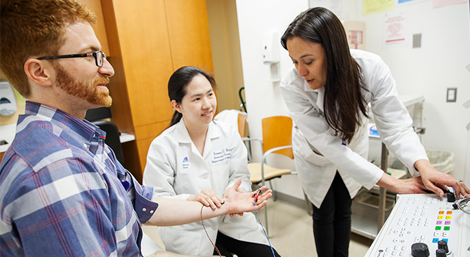
Electromyography and Nerve Conduction Studies
Electromyogram and nerve conduction studies (NCS) are tests of nerve and muscle function. They can help diagnose amyotrophic lateral sclerosis (ALS) and other motor neuron diseases, peripheral neuropathy, radiculopathy (pinched nerve) in the neck or lower back, myopathy, and neuromuscular junction disorders including myasthenia gravis.
We divide testing into two parts. First, one of our neurologists, fellows, or technicians will typically conduct a nerve conduction study. We attach small metal disc electrodes to the skin over a muscle or nerve, usually in the hand or foot, followed by another on the skin over a nearby nerve, usually on the arm or leg. We apply a small electrical current to stimulate the nerve and then record the response. Then a doctor manages the second part, a needle electromyography (EMG), which involves inserting a small needle into muscles to measure muscular activity.
What to expect
You may experience slight discomfort and minimal bleeding and bruising during this procedure, but most patients do not consider it painful. Our doctors and technicians do their best to make sure you are as comfortable as possible. The length of the procedure will depend on the number of nerves and muscles we need to test. Typically, we stimulate four to five nerves and measure five to six muscles per limb. In general, these nerve conduction studies take about 15 to 20 minutes per limb, and the EMG, about 5 to 10 minutes. A test of the right arm and leg, for example, might take an hour.
How to prepare
You do not need to make any special preparations for a nerve conduction study and you may eat and take medications on the day of your tests. We ask that you do not use lotion on your arms or legs, as it prevents the electrodes from sticking properly to your skin.
Please tell the doctor if you are taking any blood thinning medication. We will still be able to do the study, but we may need to avoid certain muscles, to reduce the risk of bleeding.
Autonomic Function Tests
The autonomic nervous system controls the function of your internal organs, such as your heart rate, blood pressure, and movement within your stomach and intestines. We may order autonomic function tests if you experience dizziness or fainting, or if we suspect you may have a small fiber neuropathy. This test, also called “small fiber testing” or “tilt table testing,” takes 90 minutes to 2 hours and is not painful.
What to Expect
Autonomic function tests come in three parts, the first is the quantitative sudomotor axon reflex test (QSART), which measures your sweat output. Sweat output may be impaired in disorders of the autonomic nervous system such as neuropathy. To perform this test, a technician attaches four small plastic capsules to your arms and legs. These capsules contain a liquid that causes sweating. The capsules are attached to a computer, which sends a small amount of electricity through the liquid and measures your sweat response. The QSART test is not dangerous or painful. You will feel a slight pricking sensation under the capsule, and your skin may look a little red after the test, but this will go away in a few hours.
The second part is cardiovagal testing, which measures how your heart rate and blood pressure respond to changes in your breathing pattern. The technician puts electrocardiogram stickers attached to wires on your chest to monitor your heart rate, and a small cuff on your finger to check your blood pressure. The cuff fills with air and squeezes your finger throughout the test. You then do two breathing exercises. The first exercise, called heart rate response to deep breathing, involves taking slow, deep breaths. The second exercise, called the Valsalva maneuver, involves blowing hard into a tube.
The final part is tilt table testing, which involves lying flat on a table with a seatbelt across your body and your feet on a footrest. After a period of rest, the table tilts up so that you are in a standing position. A technician will monitor you for 10 minutes. If you don’t feel well during this test, tell us and we will bring you down right away.
How To Prepare
In preparation for the tests, do not smoke or drink coffee, tea, or other caffeinated beverages for at least four hours beforehand. Avoid taking any medications that dry out your eyes or mouth, such as cold and allergy medications or antidepressants, for 24 hours before the test.

Evoked Potentials
Evoked potentials measure the brain’s response to a sensory stimulus. There are three main types of evoked potentials. The first is called visual evoked potentials (VEP), and it tests your visual system. We apply electrodes to your scalp then ask you to sit in front of a computer screen with one eye covered. When testing begins, a black-and-white checkerboard pattern flashes on the screen, and we ask you to watch a red dot in the middle of the screen. We repeat the procedure with your other eye covered.
The second type is somatosensory evoked potentials (SSEP), which tests the function of the entire pathway that connects nerve to spinal cord to brain. We can perform SSEPs on the arms or legs. As you lay flat, we apply electrodes to your scalp and spine, and place a stimulator on your wrist or ankle. You will feel a painless, repetitive electrical stimulus under the stimulator, and we record your brain’s response. We may repeat this procedure on the other side.
The third type is brainstem auditory evoked response, which tests the function of the pathway from your ear to auditory nerve to brain. We apply electrodes to your scalp and place headphones over your ears. When testing begins, you will hear a clicking noise in one ear. Then we test the other ear.
Skin Biopsy
A skin biopsy helps us diagnose small fiber sensory neuropathy, which is a condition that can cause pain, burning, tingling, and numbness. If the condition affects small autonomic fibers, you may experience dry mouth, dry eyes, dizziness, bowel constipation, and difficulty sweating.
Skin biopsy is a minor surgical procedure that takes about 10 minutes. We typically do this procedure at three sites in one leg: distal leg, distal thigh, and proximal thigh. The biopsied area is typically very small and you will not need sutures. The biopsy site should heal in one week and should not limit your physical activity.
Muscle and Nerve Biopsy
We typically perform muscle and nerve biopsies when we need to evaluate myopathies and neuropathies that require specific, targeted clinical treatment. Muscle biopsies help us diagnose a variety of muscle diseases, including inflammatory myopathies, metabolic myopathies, toxic myopathies, and muscular dystrophies. We perform nerve biopsies to diagnose vasculitic neuropathy, amyloid neuropathy, and demyelinating neuropathy.
We perform muscle or nerve biopsy in an operating room under monitored anesthesia with a pre-surgical evaluation two hours prior. The surgery takes about one hour.
Neuromuscular Ultrasound
Neuromuscular ultrasound uses sound waves to show us the structure of the muscles and the nerves. The test is safe and painless and does not expose your body to radiation. The images show any inflammation, trauma, or tumors of the nerves and muscles. It can also display where the muscles are so we can administer botulinum toxin injections in precise locations.