Breast Imaging
There is no more calm and welcoming place than the Blavatnik Family Chelsea Medical Center at Mount Sinai for breast imaging. We offer mammograms and other breast imaging technologies when you are concerned about a lump that you found, when your doctor recommends that you get an expert diagnosis, or when you are interested in getting a second opinion.
Our experienced breast experts use the most advanced imaging equipment in our warm, friendly setting located conveniently in the Chelsea facility. Our office is designed to enhance your comfort and privacy while we do all we can to ensure the most successful outcome possible for you.
Why Us?
Our Women’s Cancer Program breast imagers are experienced fellowship-trained specialists who treat you with compassion. We have the expertise to provide the best possible treatment and results, starting with your diagnosis.
We know that properly performing and interpreting the imaging tests is necessary for developing the correct diagnosis and most appropriate individualized treatment plan.
When to Have a Mammogram
The medical profession recommends that women have a mammogram after their 40th birthday and every year after.
If, during your annual mammogram, your doctor discovers something questionable, we welcome you to come to us at the Blavatnik Center for follow-up breast imaging.
However you are referred to us, our goal as breast specialists in the Women’s Cancer Program at the Blavatnik Center is to find any problems and treat you successfully.
We also offer the special Surveillance Breast Program for women who are at higher risk for breast cancer due to personal or family history or genetic mutation.
What is a Mammogram?
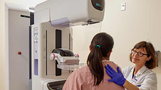
Mammograms are very-low-dose X-ray images of your breast that help us see tumors that are too small to feel. The images also allow us to detect tiny deposits of calcium that we might want to check further. Often we take two pictures of each breast so we can be certain that we see everything. The radiation dose of this testing is very low, much less than what you get from just walking around New York City.
To take the scan, our technologists compress your breast between two plastic plates. This process can feel a little awkward, but we work with you to make it as quick and as comfortable as possible. Some women ask if they can have a sonogram instead of a mammogram, but we have found that the mammogram is the most effective imaging test available to detect a tiny, early cancer.
Advanced Imaging Tests
Typically, breast monitoring starts with digital mammography, performed annually as a screening test for women over age 40, and for women with a suspected condition. If a mammogram does not provide enough information, we have a variety of other advanced imaging tests available that can tell us more. We routinely perform a variety of advanced tests:
- Breast-specific gamma imaging uses a radioactive tracer and gamma camera to pinpoint areas of cancerous tissue. We often use this as a follow-up to a mammogram for women with dense breasts.
- Computed tomography (CT) scans create detailed images of your organs, bones, and blood vessels. A fast, noninvasive approach, CT scans are highly accurate.
- Magnetic resonance imaging (MRI) tests use a magnetic field and radio wave energy to make pictures of your breast. We typically use this test to check for tumors after a breast cancer diagnosis.
- Positron emissions tomography (PET) scans use a radioactive tracer to help us identify cancer, heart disease, and brain disorders. Sometimes we combine PET scans with MRIs or CT scans. Our new machinery cuts the testing time significantly from approximately 75 minutes to about 45 minutes.
- Ultrasound is a probe that creates sound waves and produces pictures of your body’s organs, bones, and blood vessels without use of radiation. Particularly effective for dense breasts, ultrasound tests have no known harmful effects. We use ultrasound for both breasts and vaginal areas.
- X-rays are an imaging test that we use primarily for people who are being treated for advanced breast cancer, to see if the cancer has spread or if there is any lung inflammation. A mammogram is a combination of multiple X-ray tests.
Our expert breast pathologists review your imaging tests and provide detailed reports that our medical oncologists and breast surgeons use to help determine the most efficient and effective treatment approach for you.