Mammogram
Mammography; Breast cancer - mammography; Breast cancer - screening mammography; Breast lump - mammogram; Breast tomosynthesis; 3D mammography; 2D mammography; Diagnostic mammogram
A mammogram is an x-ray picture of the breasts. It is used to evaluate some breast symptoms and to find breast cancer in women with no symptoms.
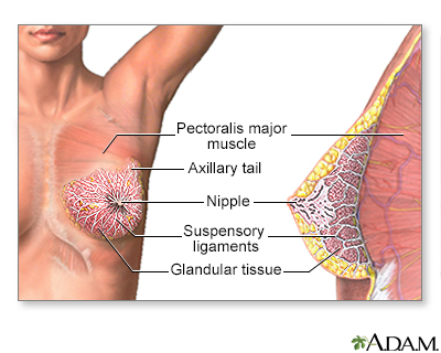
The female breast is either of two mammary glands (organs of milk secretion) on the chest.
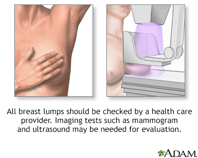
Less than one-fourth of all breast lumps are found to be cancerous, but benign breast disease can be difficult to distinguish from cancer. Consequently, all breast lumps should be checked by a health care professional.
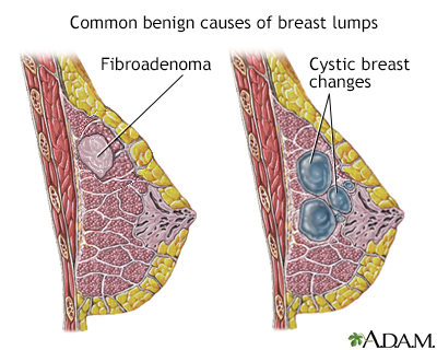
Most breast lumps are benign (non-cancerous), as in fibroadenoma, a condition that mostly affects women under age 30. Fibrocystic breast changes occur in more than 60% of all women. Fibrocystic breast cysts change in size with the menstrual cycle, whereas a lump from fibroadenoma does not. While most breast lumps are benign, it is important to identify those that are not. See your health care provider if a lump is new, persistent, growing, hard, immobile, or causing skin deformities.
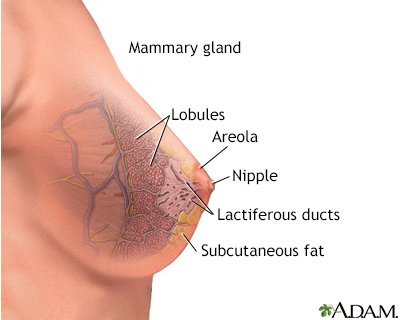
The anatomy of the breast includes the lactiferous, or milk ducts, and the mammary lobules.
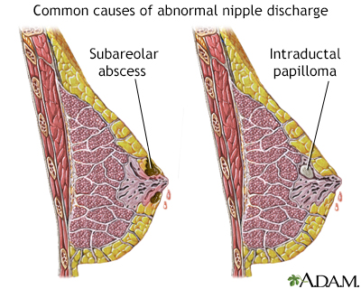
Abnormal nipple discharge may be described as any discharge not associated with lactation. The nature of the discharge may range in color, consistency and composition, and occur in one or both breasts.
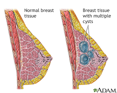
Fibrocystic breast disease is a common and benign change within the breast characterized by a dense irregular and bumpy consistency in the breast tissue. Mammography or biopsy may be needed to rule out other disorders.
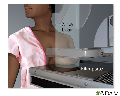
A mammogram is an x-ray picture of the breasts. It is used to find tumors and to help tell the difference between noncancerous (benign) and cancerous (malignant) disease. One breast at a time is rested on a flat surface that contains the x-ray plate. A device called a compressor is pressed firmly against the breast to help flatten out the breast tissue. Each breast is compressed horizontally, then obliquely and an x-ray is taken of each position.
How the Test is Performed
You will be asked to undress from the waist up. You will be given a gown to wear. Depending on the type of equipment used and your physical condition, you will sit or stand.
One breast at a time is rested on a flat surface that contains the x-ray plate. A device called a compressor will be pressed firmly against the breast to help flatten the breast tissue. This enables the radiologist to make a better evaluation of your breasts.
The x-ray pictures are taken from several angles. You may be asked to hold your breath as each picture is taken.
You may be asked to come back at a later date for more mammogram images. This does not always mean you have breast cancer. Your health care provider may simply need to recheck an area that could not be clearly seen on the first test.
TYPES OF MAMMOGRAPHY
Traditional mammography uses film, similar to other x-rays.
Digital mammography is the most common technique:
- It is now used in most breast screening centers.
- It allows the x-ray image of the breast to be viewed and manipulated on a computer screen.
- It may be more accurate in younger women with dense breasts. It has not yet been proven to help reduce a woman's risk of dying of breast cancer compared to film mammography.
Three-dimensional (3D) mammography is a type of digital mammography.
How to Prepare for the Test
DO NOT use deodorant, perfume, powders, or ointments under your arms or on your breasts on the day of the mammogram. These substances may hide a portion of the images or produce an artifact that can falsely look like an abnormality. Remove all jewelry from your neck and chest area.
Tell your provider and the x-ray technologist if you are pregnant or breastfeeding, or if you've had a breast biopsy.
How the Test will Feel
The compressor surfaces may feel cold. When the breast is pressed down, you may have some pain. This needs to be done to get good quality images.
Why the Test is Performed
When and how often to have a screening mammogram is a choice you must make. Different expert groups do not fully agree on the best timing for this test.
Before having a mammogram, talk to your provider about the pros and cons of having the test. Ask about:
- Your risk for breast cancer
- Whether screening decreases your chance of dying from breast cancer
- Whether there is any harm from breast cancer screening, such as side effects from testing or overtreatment of cancer when it's discovered
Mammography is performed to screen women to detect early breast cancer when it is more likely to be cured. Mammography is generally recommended for:
- Women starting at age 40, repeated every 1 to 2 years. (This is not recommended by all expert organizations.)
- All women starting at age 50, repeated every 1 to 2 years.
- Women with a mother or sister who had breast cancer at a younger age should consider yearly mammograms. They should begin earlier than the age at which their youngest family member was diagnosed.
Mammography is also used to:
- Follow a woman who has had an abnormal mammogram.
- Evaluate a woman who has symptoms of a breast disease. These symptoms may include a lump, nipple discharge, breast pain, dimpling of the skin on the breast, changes of the nipple, or other findings.
Of all the different types of cancers, breast cancer is one of the most talked about, and with good reason. One out of every eight women will develop breast cancer sometime in their life. That's why every woman should be thinking about how to protect herself from this disease. Breast cancer is cancer that forms in the breast. Usually, it begins in the tubes that transport milk from the breast to the nipple. If the cancer spreads to other parts of the breast or body, it's called invasive breast cancer. Some breast cancers are more aggressive, growing more quickly than others. Although women are 100 times more likely to develop breast cancer, men can also get the disease because they do have breast tissue. You're more likely to get breast cancer if you're over 50, you started your periods before age 12, or you have a close family member with the disease. Drinking more than a couple of glasses of alcohol a day and using hormone replacement therapy for several years also may increase your risk. The telltale sign of breast cancer is a lump in your breast or armpit. You may also notice a change in the shape, size, or texture of your breast, or have fluid coming from your nipple when you're not breastfeeding. If you notice any changes in your breasts, call your doctor. You'll probably need to have an imaging scan, such as a mammogram, MRI, or ultrasound. A piece of tissue may be removed from your breast, called a biopsy. With these tests, your doctor can tell whether you have breast cancer, and if so, determine whether or not it has spread. So, how do we treat breast cancer? That really depends on the type of cancer, and how quickly it's spreading. Your doctor may recommend that you have the cancer removed with surgery. Sometimes it's enough just to remove the lump. That's called a lumpectomy. In other cases, the doctor will need to remove the entire breast to get rid of all the cancer or prevent it from coming back. That's called a mastectomy. Other treatments for breast cancer include chemotherapy, medicines that kill cancer cells, and radiation therapy, which uses energy to destroy cancer. Women whose cancer is fueled by the hormone estrogen may receive hormone therapy to block the effects of estrogen on their cancer. Today's breast cancer treatments are better than ever. Many women who have breast cancer go on to live long, healthy lives. The outlook really depends on how fast the tumor is growing, and how far it has spread. That's why it's so important to report any changes in your breasts to your doctor as soon as you notice them. Women who are at an especially high risk for breast cancer because of their family history can talk to their doctor about taking medicine or even having surgery to reduce their risk.
Normal Results
Breast tissue that shows no signs of a mass or suspicious looking calcifications is considered normal.
What Abnormal Results Mean
Most abnormal findings on a screening mammogram turn out to be benign (not cancer) or nothing to worry about. New findings or changes must be further evaluated.
A radiology doctor (radiologist) may see the following types of findings on a mammogram:
- A well-outlined, smoothly marginated, rounded spot (this is more likely to be a noncancerous condition, such as a cyst)
- Irregular appearing masses or lumps
- Dense areas in the breast that can be breast cancer or hide breast cancer
- Calcifications, which are caused by tiny deposits of calcium in the breast tissue (most calcifications are not a sign of cancer)
At times, the following tests are also needed to further assess mammogram findings:
- Additional mammogram views, including magnification or compression views
- Breast ultrasound
- Breast MRI exam (less commonly done)
Comparing your current mammogram to your past mammograms helps the radiologist tell whether you had an abnormal finding in the past and whether it has changed.
When mammogram or ultrasound results look suspicious, a biopsy is done to test the tissue and see if it is cancerous. Types of biopsies include:
- Stereotactic (guided)
- Ultrasound (guided)
- Open (surgical)
Risks
The level of radiation is low and any risk from mammography is very low. If you are pregnant and need to have an abnormality checked, your belly area will be covered and protected by a lead apron.
Routine screening mammography is not done during pregnancy or while breastfeeding.
References
American Cancer Society website. American Cancer Society recommendations for the early detection of breast cancer.
American College of Obstetricians and Gynecologists (ACOG) website. ACOG Practice Bulletin: Breast cancer risk assessment and screening in average-risk women. No. 179, July 2017.
Henry NL, Shah PD, Haider I, Freer PE, Jagsi R, Sabel MS. Cancer of the breast. In: Niederhuber JE, Armitage JO, Kastan MB, Doroshow JH, Tepper JE, eds. Abeloff's Clinical Oncology. 6th ed. Philadelphia, PA: Elsevier; 2020:chap 88.
National Cancer Institute website. Breast cancer screening (PDQ) - health professional version.
Siu AL; US Preventive Services Task Force. Screening for breast cancer: US Preventive Services Task Force recommendation statement. Ann Intern Med. 2016;164(4):279-296. PMID: 26757170
Version Info
Last reviewed on: 1/14/2023
Reviewed by: Neil Grossman, MD, MetroWest Radiology Associates, Framingham, MA. Review provided by VeriMed Healthcare Network. Also reviewed by David C. Dugdale, MD, Medical Director, Brenda Conaway, Editorial Director, and the A.D.A.M. Editorial team.
